Insights

Intrinsic Imaging's Guidance for Sponsors

Guidance for Sponsors from Intrinsic Imaging physicians on therapeutic areas and indications, imaging endpoints, imaging core lab methods and clinical trial design.

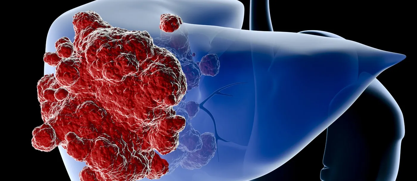
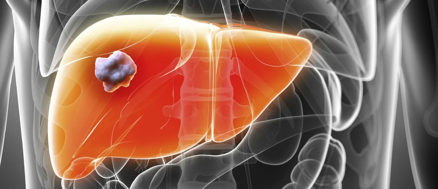
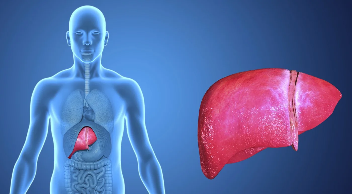
Illustration of the human liver

Guidance for Sponsors · Volume 4, Issue 1 · 2026 Edition

Magnetic Resonance Elastography: A Quantitative Endpoint for Liver Fibrosis Trials

Magnetic resonance elastography (MRE) measures liver stiffness without a needle, and it has become one of the most accurate imaging tools available for staging liver fibrosis. Now that the FDA has approved the first therapies for metabolic dysfunction-associated steatohepatitis (MASH), sponsors developing the next generation of liver therapies need endpoints that are quantitative, repeatable and acceptable to patients. Combined with MRI proton density fat fraction (MRI-PDFF) in a single liver MRI exam, MRE provides exactly that: a one-minute acquisition that stages fibrosis and tracks how it changes over the course of a trial.

Read the article
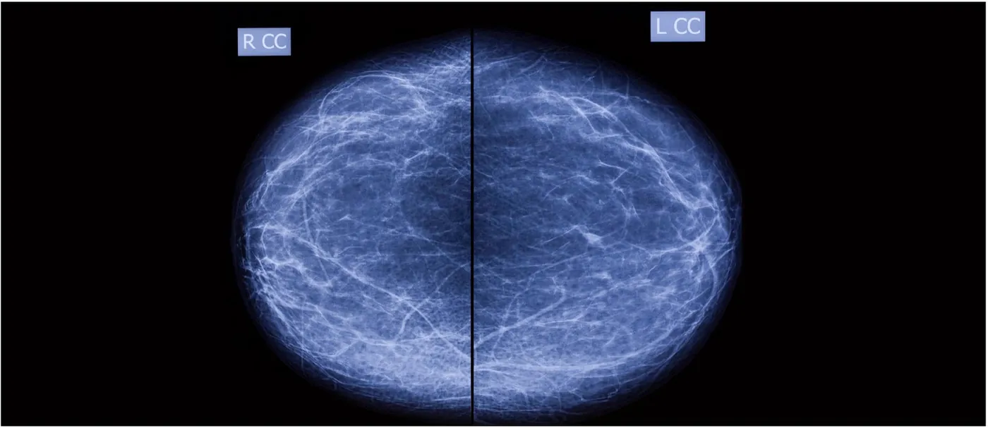
Bilateral craniocaudal screening mammogram

Guidance for Sponsors · Volume 4, Issue 2 · 2026 Edition

Screening Mammography AI: A Continued Market Opportunity

Computer-aided detection (CAD) promised to help radiologists find more breast cancers, but a decade of large studies showed that first-generation systems added false alarms without improving accuracy. Artificial intelligence has changed that picture. Randomized and nationwide studies published since 2023 show AI-supported screening detecting more cancers, reducing interval cancers and cutting radiologist workload without raising recall rates. For developers, the opportunity is larger than ever. So is the burden of proof: regulators, payers and screening programs now expect rigorous reader studies and real-world evidence.

Read the article
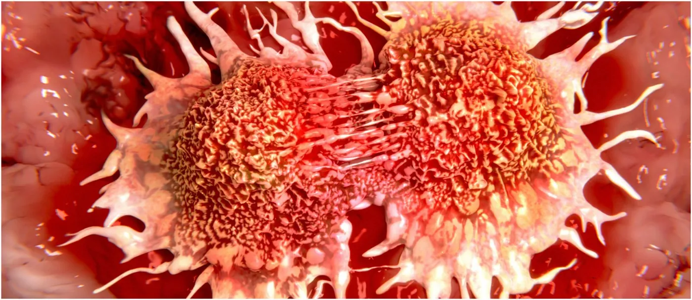
Illustration of dividing cancer cells

Guidance for Sponsors · Volume 4, Issue 3 · 2026 Edition

Oncologic Imaging: The Benefits of Diffusion-Weighted MRI

Diffusion-weighted MRI (DWI) was first used to greatest advantage in stroke imaging. Advances in gradients and pulse sequences have since made it a core tool for detecting, characterizing and following malignant lesions in the chest, abdomen and pelvis. Today it is the dominant sequence for peripheral-zone prostate cancer, a foundation of whole-body MRI for bone and marrow disease, and, through the apparent diffusion coefficient (ADC), a quantitative biomarker with published repeatability standards. For sponsors, DWI offers a fast, contrast-free measure of tumor cellularity and treatment response, provided acquisition and analysis are standardized across sites.

Read the article
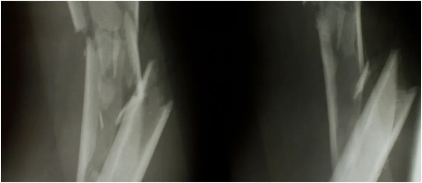
Radiographs of a comminuted long-bone diaphyseal fracture

Guidance for Sponsors · Volume 4, Issue 4 · 2026 Edition

Imaging Analysis and Operative Treatment of Diaphyseal Fractures

Fractures are among the most common reasons for an imaging examination, and imaging guides every stage of care: diagnosis, classification, reduction, fixation, detection of complications and confirmation of healing. Radiography, CT, MRI and bone scintigraphy each play a distinct role. For sponsors of orthopedic drug, biologic and device trials, the central challenge is that fracture union, the most commonly reported primary outcome, still has no universally accepted radiographic definition. Standardized scoring, consistent imaging schedules and independent central review turn a subjective judgment into a defensible endpoint.

Read the article
Illustration of the human spine and skeleton

Guidance for Sponsors · Volume 4, Issue 5 · 2026 Edition

Orthopedic Device Assessments: Medical Imaging Challenges in Clinical Trials

Orthopedic research spans joint reconstruction, spinal fusion and motion preservation, and biologics for cartilage, tendon and ligament repair. Regulators and payers expect rigorous quantitative and qualitative evidence of efficacy, safety and longevity, and imaging supplies much of it. Because investigational devices are often unlike anything imaged before, protocols must be built around the device itself: its engineering, materials, surgical technique and position. Advances in CT, metal-artifact-reduction MRI and radiostereometric analysis now allow earlier and more precise answers than ever.

Read the article
Illustration of the lungs and airways

Guidance for Sponsors · Volume 4, Issue 6 · 2026 Edition

The Challenges of Reducing Lung Cancer Mortality with Screening

Lung cancer remains the leading cause of cancer death in the United States. Two large randomized trials, NLST and NELSON, have shown that low-dose CT (LDCT) screening reduces lung cancer mortality, and U.S. eligibility was broadened in 2021. Yet the challenges that shaped the first edition of this issue remain: false positives, overdiagnosis, radiation, and the biases that can make screening look more effective than it is. Artificial intelligence for nodule detection and characterization adds new opportunity, and new questions that only well-designed trials can answer.

Read the article
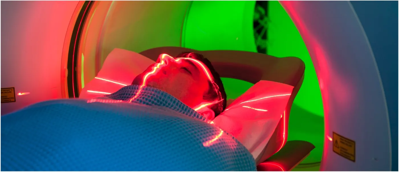
Patient positioned in a CT scanner

Guidance for Sponsors · Volume 4, Issue 7 · 2026 Edition

Computed Tomography Data Acquisition in Clinical Trials

CT is one of the most widely used modalities in clinical trials, and a substantial literature, led by RECIST 1.1, governs how lesions are measured. Far less attention is paid to how the images are acquired, yet patient motion, scanner variability, slice thickness and contrast timing all add error to every measurement. FDA's 2018 guidance on imaging endpoint process standards makes acquisition a regulatory concern. A few basic principles, written into the protocol and imaging charter and applied consistently at every site, substantially improve data accuracy.

Read the article
Illustration of an inflamed knee joint

Guidance for Sponsors · Volume 4, Issue 8 · 2026 Edition

Advances in High-Field MRI for the Assessment of Arthritis

Arthritis is a leading cause of disability, affecting about 53 million U.S. adults. Its central feature is loss of hyaline cartilage, which has little capacity to repair. Radiographs show only late damage; high-field 3.0 Tesla MRI shows cartilage morphology in fine detail, and compositional techniques such as T2 mapping reveal degeneration before visible loss. For sponsors, the challenge is regulatory as well as technical: FDA has not yet accepted a structural endpoint for osteoarthritis approval, so imaging must be precise, reproducible and linked to how patients feel and function.

Read the article
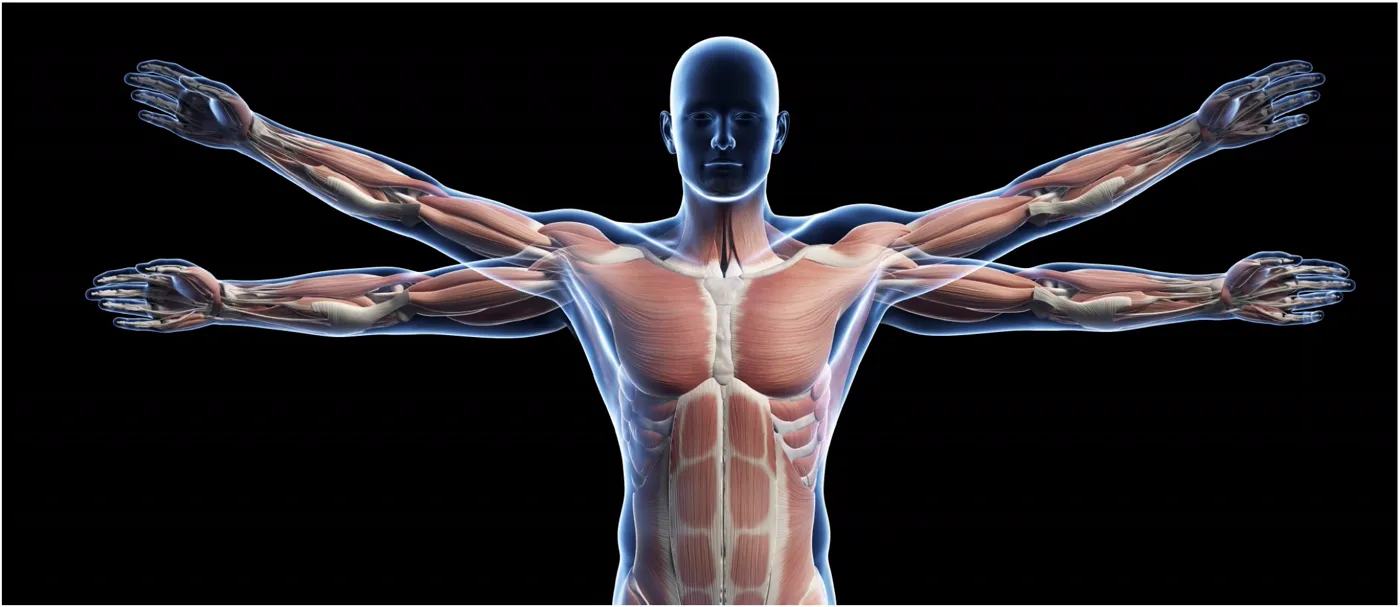
Illustration of the human muscular system

Guidance for Sponsors · Volume 4, Issue 9 · 2026 Edition

Muscular Dystrophy: Medical Imaging in Clinical Trials

MRI has become one of the most valuable tools for measuring disease in muscular dystrophy. It shows which muscles are involved, how severely and how actively, and it can guide biopsy and monitor therapy. Since the first edition of this issue, quantitative MRI has moved from grading scales to precise fat-fraction measurement, and muscle fat fraction has been shown to predict loss of ambulation in Duchenne muscular dystrophy. With new therapies now approved, MRI is a key secondary endpoint and a candidate surrogate for future trials.

Read the article
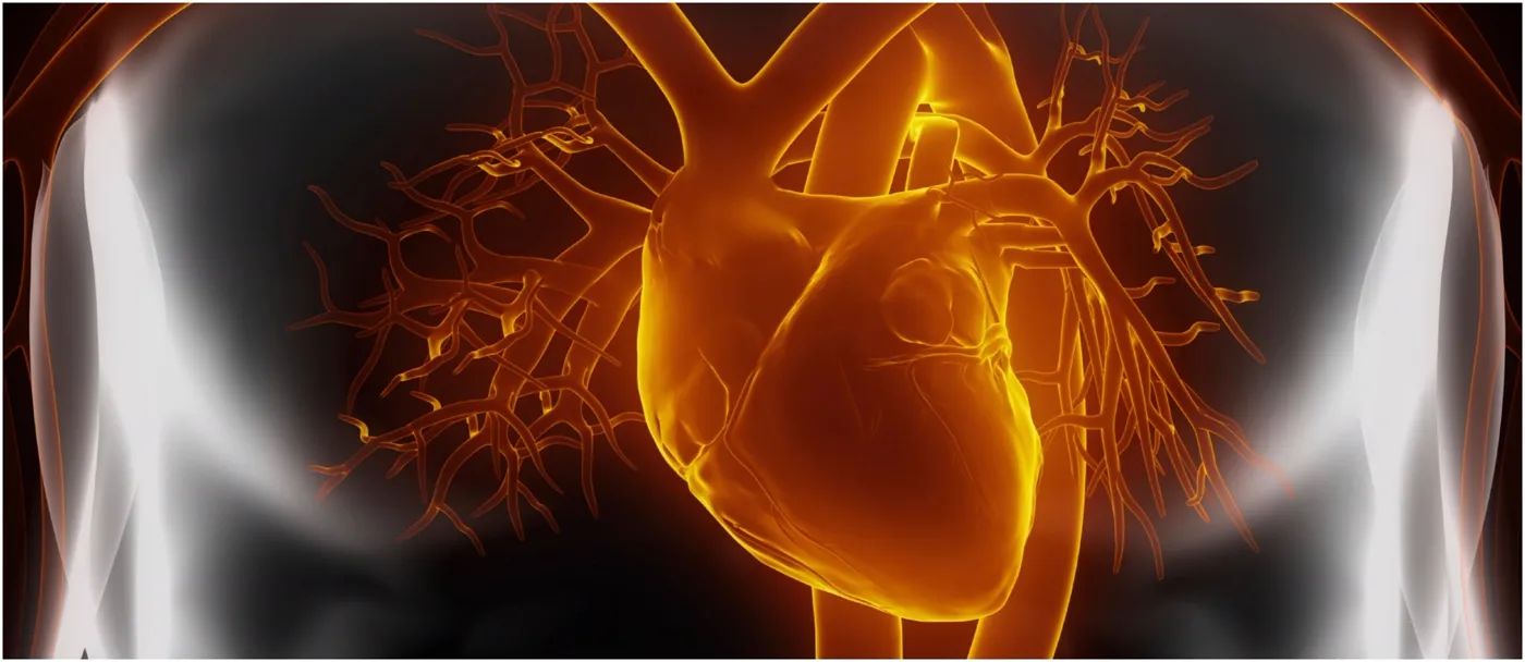
Illustration of the heart and coronary vessels

Guidance for Sponsors · Volume 4, Issue 10 · 2026 Edition

Echocardiography: Advantages in Clinical Trials

Echocardiography is safe, portable, affordable and available worldwide, which makes it one of the most practical imaging tools for clinical trials. It measures chamber size, function, valves and mass, and changes in these measures predict major cardiovascular events, allowing smaller trials and earlier answers. Since the first edition of this issue, global longitudinal strain and three-dimensional echocardiography have become standard measures, and cardio-oncology guidelines now define cardiac injury from cancer therapy partly by strain. Standardized acquisition and core-lab reading remain essential.

Read the article
Illustration of a vertebral compression fracture

Guidance for Sponsors · Volume 4, Issue 11 · 2026 Edition

Osteoporosis Therapy: Challenges in Monitoring Efficacy in Clinical Trials

Osteoporotic fractures are devastating, and proving that a new therapy prevents them has traditionally required large, long fracture trials. That changed in December 2025, when FDA qualified the 24-month change in total hip bone mineral density (BMD), measured by DXA, as a validated surrogate endpoint for phase 3 trials in postmenopausal osteoporosis. The decision makes imaging quality more important than ever. DXA must be calibrated and centrally reviewed, while QCT, vertebral fracture assessment and microarchitecture imaging add information that BMD alone cannot provide.

Read the article
Illustration of tumor cells

Guidance for Sponsors · Volume 5, Issue 1 · 2026 Edition

Immunotherapy and Imaging of Metastatic Pancreatic Adenocarcinoma

Pancreatic adenocarcinoma remains one of the most lethal cancers, with a five-year relative survival of 13% and a rising share of U.S. cancer deaths. After a decade in which checkpoint immunotherapy largely failed in this disease, the therapeutic landscape is finally changing: a new first-line chemotherapy regimen has been approved, and in 2026 an oral RAS inhibitor doubled overall survival in a Phase 3 trial of previously treated patients. These advances raise the stakes for imaging. Contrast-enhanced CT, MRI and FDG-PET/CT must deliver staging and response data that are standardized, reproducible and interpretable under the right criteria, including iRECIST when immunotherapy is involved.

Read the article
Illustration of the lungs and airways

Guidance for Sponsors · Volume 5, Issue 2 · 2026 Edition

Advances in Imaging of Non-Small Cell Lung Cancer (NSCLC)

Non-small cell lung cancer has become the model for precision oncology. Targeted therapies against EGFR, ALK and KRAS, and immunotherapy given before and after surgery, have transformed outcomes for patients who once had few options. Each advance places new demands on imaging. Tumors treated with targeted agents and immunotherapy do not always shrink in the way RECIST expects, measurement variability can rival the treatment effect, and functional and volumetric techniques are maturing as candidate biomarkers. This issue reviews how imaging assessment of NSCLC is evolving and what sponsors must get right.

Read the article
Illustration of a neuron

Guidance for Sponsors · Volume 5, Issue 3 · 2026 Edition

Functional Imaging of the Dopamine Pathway

Parkinson disease is rising rapidly worldwide, and its early diagnosis remains difficult: symptoms overlap with essential tremor, drug-induced parkinsonism and the atypical parkinsonian syndromes, and routine MRI is usually normal. Functional imaging of the presynaptic dopamine pathway, with DaTscan SPECT and Fluorodopa F 18 PET, makes the loss of dopaminergic neurons visible. European regulators have qualified dopamine transporter imaging as an enrichment biomarker for early Parkinson disease trials, and it now works alongside the alpha-synuclein seed amplification assay in a new biological definition of the disease. This issue explains how these tools work and how sponsors should use them.

Read the article
Illustration of neurons

Guidance for Sponsors · Volume 5, Issue 4 · 2026 Edition

Temporal Lobe Epilepsy and Volumetric MRI in Clinical Trials

Temporal lobe epilepsy caused by hippocampal sclerosis is one of the most common forms of drug-resistant focal epilepsy, and one of the most treatable when it is correctly identified. Visual reading of MRI can miss mild or bilateral hippocampal atrophy, which makes patient selection the weak link in many trials. Quantitative volumetry with FDA-cleared software, acquired on a harmonized epilepsy protocol, converts a subjective impression into a numerical biomarker. It can set inclusion thresholds, stratify severity, predict surgical outcome and, potentially, measure the effect of new drugs, ablation and neurostimulation devices.

Read the article
Illustration of an immune cell engaging a tumor cell

Guidance for Sponsors · Volume 5, Issue 5 · 2026 Edition

irRECIST and Immuno-Oncology in Clinical Trials

Immunotherapy has changed the practice of oncology, and it has changed the practice of measuring response. Tumors treated with checkpoint inhibitors can enlarge, or show new lesions, before they shrink, a pattern that conventional criteria classify as failure. Over fifteen years, response assessment has moved from the immune-related response criteria of 2009, through unidimensional irRC and irRECIST, to iRECIST, the RECIST Working Group's consensus standard. This issue traces that evolution, compares the criteria and explains how sponsors should apply them so that a working therapy is not mistaken for a failing one.

Read the article
Illustration of the heart and blue blood vessels

Guidance for Sponsors · Volume 5, Issue 6 · 2026 Edition

Contrast Echocardiographic Imaging in Clinical Trials

Ultrasound enhancing agents turn a difficult echocardiogram into a diagnostic one. By opacifying the left ventricle and sharpening the endocardial border, they improve the accuracy and reproducibility of ejection fraction and wall motion analysis, measures that sit at the heart of many cardiovascular and cardio-oncology trials. Over the past two decades, regulators have progressively relaxed the restrictions placed on these agents as their safety record has grown. This issue reviews the technology, its regulatory history and the role of myocardial perfusion, and sets out what sponsors should specify when contrast echocardiography supports a trial endpoint.

Read the article
Illustration of the heart and coronary arteries

Guidance for Sponsors · Volume 5, Issue 7 · 2026 Edition

Coronary Artery Calcium Scoring in Clinical Trials

Coronary artery calcium scoring is one of the most powerful and economical predictors of cardiovascular risk, and an established biomarker in trials of preventive therapy. Yet the score is less simple than it appears. Calcium density carries prognostic information of its own, scores vary between scanners, vendors and software, and photon-counting CT is now changing the measurement itself. This issue reviews the method, the CAC-DRS reporting system and the sources of variability that sponsors must control when calcium scoring supports a trial endpoint.

Read the article
Illustration of a brain tumor within the head

Guidance for Sponsors · Volume 5, Issue 8 · 2026 Edition

The Challenge of Quantitative Measurements of Primary Brain Tumors in Clinical Trials

Glioblastoma remains the most common and most lethal malignant primary brain tumor, with a median survival of nine months. Measuring its response to treatment is uniquely difficult: gliomas infiltrate normal brain, contain necrosis by definition, and are mimicked by treatment effects that enhance and grow. RANO 2.0, published in 2023, unified response criteria for all gliomas, and a standardized brain tumor imaging protocol now underpins multicenter trials. The 2024 approval of vorasidenib, based on centrally reviewed radiographic progression, shows how much rests on getting imaging right. This issue explains the challenges and how sponsors can meet them.

Read the article
Illustration of an inflamed knee joint

Guidance for Sponsors · Volume 5, Issue 9 · 2026 Edition

Medical Imaging of Osteoarthritis of the Knee in Clinical Trials

Osteoarthritis of the knee is among the most common causes of disability, and among the most difficult conditions in which to prove that a therapy modifies disease. Radiographic joint space narrowing, the traditional structural endpoint, is slow, indirect and sensitive to positioning. MRI shows cartilage, bone marrow, menisci and synovium directly, and validated semiquantitative scores, quantitative cartilage morphometry and compositional mapping now offer far more sensitive measures of change. This issue reviews the strengths and limitations of each modality and how sponsors should combine them in trials of disease-modifying therapies.

Read the article
Illustration of a blood cancer cell

Guidance for Sponsors · Volume 5, Issue 10 · 2026 Edition

Medical Imaging of Chronic Lymphocytic Leukemia

Chronic lymphocytic leukemia has been transformed by targeted therapy. BTK inhibitors, BCL-2 inhibition and cellular therapies have pushed five-year survival above 90%, and in October 2026 FDA approved pirtobrutinib for previously untreated disease. Imaging plays a narrower but critical role in this disease: international guidelines do not require CT in routine practice, yet they consider it desirable in clinical trials, and FDG-PET/CT is the key tool for detecting Richter transformation. This issue sets out where imaging belongs in a CLL trial and how to standardize it.

Read the article
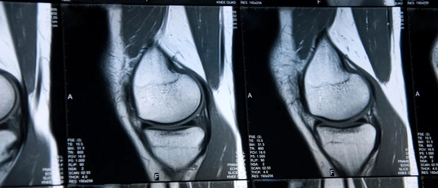
MRI image of the knee

Guidance for Sponsors · Volume 5, Issue 11 · 2026 Edition

Quantitative MRI Imaging in Orthopedic Clinical Trials

Orthopedic trials have long depended on patient questionnaires and on radiographic joint space width, a measurement of what cannot be seen. Quantitative MRI measures cartilage, bone and soft tissue directly, in three dimensions, and can reveal biochemical change before structural loss occurs. Validated scoring systems for rheumatoid arthritis, consensus profiles for compositional cartilage imaging and metal artifact reduction techniques for implants have made MRI a practical endpoint across arthritis and device trials. This issue reviews those methods and what sponsors must standardize to use them well.

Read the article
Digital illustration of a human head in profile with glowing neural network connections representing quantitative brain imaging

Guidance for Sponsors · Volume 6, Issue 1 · 2026 Edition

Quantitative Neuroimaging of Alzheimer's Disease

An estimated 7.2 million Americans aged 65 and older are living with Alzheimer's dementia in 2025, at a cost to the nation of about $384 billion.[1] For most of the past century the diagnosis was clinical and could only be confirmed at autopsy. That has changed. Amyloid PET, tau PET, volumetric MRI and, since 2025, FDA-cleared blood tests now define the disease biologically while a patient is alive, and the first disease-modifying antibodies are approved. Quantitative neuroimaging has moved from a research tool to the backbone of how Alzheimer's trials enroll, treat and monitor patients. This issue explains the imaging biomarkers sponsors must plan for.

Read the article
Translucent illustration of a human head with the brain highlighted in red to represent traumatic brain injury

Guidance for Sponsors · Volume 6, Issue 2 · 2026 Edition

Traumatic Brain Injury: Medical Imaging in Clinical Trials

Traumatic brain injury (TBI) caused about 69,000 deaths in the United States in 2021 and more than 214,000 hospitalizations in 2020, and an estimated 69 million people worldwide sustain a TBI each year.[1,2] Despite decades of effort, no neuroprotective drug has succeeded in a Phase 3 trial, and the heterogeneity of the injury is a leading explanation. Imaging is central to solving that problem. CT remains the first-line tool, MRI reveals injury CT cannot see, blood biomarkers now help decide who needs a scan, and a new classification framework published in 2025 makes imaging a formal pillar of how TBI is characterized. This issue reviews what sponsors need to know.

Read the article
Illustration of a liver with a cluster of red tumor cells on its surface, representing imaging of tumor response

Guidance for Sponsors · Volume 6, Issue 3 · 2026 Edition

Imaging and Monitoring Tumor Response in Clinical Trials

Imaging is the way most cancer trials measure whether a drug works. Objective response rate, progression-free survival and duration of response all depend on images acquired at protocol-defined intervals and interpreted by consistent rules. Over the past fifteen years those rules have multiplied: RECIST 1.1 remains the default, but immunotherapy, molecularly targeted agents, locoregional therapies and PET have each produced their own criteria. This issue explains the current landscape of response assessment and what sponsors must build into a protocol to make imaging endpoints hold up to regulatory review.

Read the article
Translucent illustration of the human torso from behind showing the spine and rib cage

Guidance for Sponsors · Volume 6, Issue 4 · 2026 Edition

Complex Spinal Deformities: Medical Imaging in Clinical Trials

Scoliosis affects two to three percent of adolescents, and degenerative adult spinal deformity is far more common than once thought, with radiographic prevalence reported as high as 68 percent in volunteers over 60.[1] Deformity surgery has become one of the most complex and heavily instrumented fields in orthopedics, and the devices, growth-modulating implants and surgical strategies it uses are tested in trials whose primary endpoints are radiographic. This issue reviews how spinal deformity is classified and measured, how imaging technology has changed, and what sponsors need from an imaging core lab.

Read the article
Illustration of a human skeleton with the hand and wrist joints glowing to indicate joint inflammation

Guidance for Sponsors · Volume 6, Issue 5 · 2026 Edition

Advances in Imaging of Rheumatoid Arthritis

Rheumatoid arthritis (RA) affected an estimated 17.6 million people worldwide in 2020, and that number is projected to approach 32 million by 2050.[1] Biologic and targeted synthetic disease-modifying drugs have transformed outcomes, and every one of them reached the market with imaging evidence that it slows structural joint damage. Radiography remains the regulatory standard for that claim, but MRI and ultrasound detect inflammation and erosion months before radiographs change, and standardized scoring systems now exist for all three modalities. This issue reviews how imaging is used in RA trials and what a decade of imaging-guided treatment studies has taught sponsors.

Read the article
Illustration of the human torso with the heart, lungs and liver highlighted in warm colors

Guidance for Sponsors · Volume 6, Issue 6 · 2026 Edition

Tissue Iron Deposition: MRI Quantitation in Clinical Trials

Iron overload is the inevitable consequence of chronic transfusion and of ineffective erythropoiesis in thalassemia, sickle cell disease and myelodysplastic syndromes. Each unit of blood delivers about 200 mg of iron the body cannot excrete, and the resulting deposition in liver, heart and endocrine organs was for decades the leading cause of death in transfusion-dependent thalassemia. MRI changed that. Non-invasive measurement of liver iron concentration and cardiac T2* replaced biopsy, revealed cardiac iron that serum ferritin could not predict and gave chelation trials objective endpoints. With gene therapies and new disease-modifying drugs now approved, quantitative iron MRI is more important than ever. This issue explains the methods and what sponsors should specify.

Read the article
Translucent illustration of a human head and shoulders with the brain and spinal nerves highlighted

Guidance for Sponsors · Volume 6, Issue 7 · 2026 Edition

Neurological Imaging: Contrast Enhancement in Clinical Trials

Intravenous contrast enhancement is one of the most informative signals in neurological imaging. Where the blood-brain barrier is intact, gadolinium stays in the vessels and the brain does not enhance; where disease has disrupted it, in tumors, active demyelination, infection or inflammation, contrast leaks into tissue and lights up the lesion. That simple principle underlies the primary endpoints of trials in glioma, multiple sclerosis and many other CNS conditions. The past decade has also brought a new understanding of gadolinium retention, a shift to macrocyclic agents, and the approval of high-relaxivity agents that work at a fraction of the traditional dose. This issue reviews the physiology, the patterns and the practical choices sponsors now face.

Read the article
Illustration of the colon with a red tumor mass in the sigmoid region representing colorectal cancer

Guidance for Sponsors · Volume 6, Issue 8 · 2026 Edition

Metastatic Colorectal Cancer and Locoregional Therapies

The American Cancer Society estimates about 158,850 new cases of colorectal cancer and 55,230 deaths in the United States in 2026, and the disease is now the leading cause of cancer death in adults under 50.[1] Roughly half of patients develop liver metastases, and for most of them the liver is what determines survival. Over the past decade the options for treating those metastases have multiplied: systemic therapy guided by molecular profiling, radioembolization, chemoembolization with drug-eluting beads, thermal ablation and, remarkably, liver transplantation. Each produces imaging changes that conventional size-based criteria handle poorly. This issue reviews the current evidence and what sponsors should build into trials of liver-directed therapy.

Read the article
Illustration of the liver with a white tumor on its surface against a dark background

Guidance for Sponsors · Volume 6, Issue 9 · 2026 Edition

WHO, RECIST, EASL, mRECIST and Cheson: Challenges of Evaluating Tumor Response

More than 100 therapeutic antibodies and a steady stream of small molecules, antibody-drug conjugates and immunotherapies have reached the market since the first monoclonal antibody was approved in 1986.[1] Nearly every one of them was judged, at least in part, by how tumors on imaging responded. The criteria used to make that judgment have evolved from the WHO handbook of 1981 through RECIST, its 2009 revision, immune-specific criteria, liver-specific criteria for hepatocellular carcinoma and the PET-based Lugano classification for lymphoma. Each was created to fix a weakness in its predecessor, and each has limitations of its own. This issue traces that evolution and offers practical guidance on choosing and applying criteria in a trial.

Read the article

Illustration of the human torso showing the heart and major blood vessels in red and blue

Guidance for Sponsors · Volume 6, Issue 10 · 2026 Edition

Inferior Vena Cava Filters: Medical Imaging in Clinical Trials

Venous thromboembolism affects as many as 900,000 Americans each year and causes an estimated 60,000 to 100,000 deaths.[1] Anticoagulation is the treatment, but for patients who cannot be anticoagulated, an inferior vena cava (IVC) filter offers mechanical protection against pulmonary embolism. Filter use expanded dramatically with the introduction of retrievable devices, then came under scrutiny when the FDA warned of fractures, migration and perforation and when randomized evidence failed to show benefit in patients who could receive anticoagulation. Since then a large prospective trial has defined the safety of modern filters, professional societies have issued evidence-based guidelines and imaging has become central to both the indication for a filter and its follow-up. This issue reviews the evidence and the imaging a filter trial requires.

Read the article
Request a consultation

Tell us what you are planning and our team will follow up to discuss your upcoming trial.