
Guidance for Sponsors · Volume 6, Issue 9 · 2026 Edition
WHO, RECIST, EASL, mRECIST and Cheson: Challenges of Evaluating Tumor Response
By Patrick Middlebrook, M.D., Diagnostic Radiology · Updated for 2026 by Intrinsic Imaging
Download the PDF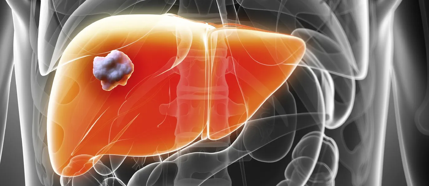
More than 100 therapeutic antibodies and a steady stream of small molecules, antibody-drug conjugates and immunotherapies have reached the market since the first monoclonal antibody was approved in 1986.[1] Nearly every one of them was judged, at least in part, by how tumors on imaging responded. The criteria used to make that judgment have evolved from the WHO handbook of 1981 through RECIST, its 2009 revision, immune-specific criteria, liver-specific criteria for hepatocellular carcinoma and the PET-based Lugano classification for lymphoma. Each was created to fix a weakness in its predecessor, and each has limitations of its own. This issue traces that evolution and offers practical guidance on choosing and applying criteria in a trial.
WHO to RECIST: Simplifying Measurement
The WHO criteria of 1981 were the first widely adopted standard. They measured lesions bidimensionally, summed the products of perpendicular diameters and defined partial response as a 50 percent decrease and progression as a 25 percent increase. They left many details, such as how many lesions to measure and what counted as measurable, to the investigator, and results diverged across trials.
RECIST, published in 2000, replaced bidimensional with unidimensional measurement, set a 30 percent decrease for partial response and a 20 percent increase for progression, and fixed a minimum lesion size. RECIST 1.1 in 2009 reduced the number of target lesions from ten to five (two per organ), defined lymph node measurement on the short axis with a 15 mm threshold for target status, required a 5 mm absolute increase in addition to the 20 percent for progression, and clarified the handling of new lesions and FDG-PET.[2] It remains the default standard for solid tumor trials and the one regulators expect unless there is a scientific reason to depart from it.[3]
Immunotherapy and iRECIST
Checkpoint inhibitors exposed a weakness: tumors can enlarge or new lesions can appear through immune infiltration before responding. The immune-related response criteria (irRC) of 2009 and later irRECIST attempted to address this using total tumor burden, but their definitions varied between trials. The RECIST Working Group published iRECIST in 2017 to provide a single, consistent framework. Its key feature is the category of unconfirmed progressive disease (iUPD), which must be confirmed on a repeat scan at least four weeks later before progression is declared, and which allows a patient who subsequently responds to be reset.[4] iRECIST is intended for exploratory endpoints and for guiding treatment beyond progression within trials; RECIST 1.1 still typically governs the regulatory primary endpoint.
Hepatocellular Carcinoma: EASL and mRECIST
Hepatocellular carcinoma (HCC) is treated largely with therapies that kill tumor without shrinking it: chemoembolization, radioembolization, ablation and antiangiogenic drugs. Size criteria therefore understate their effect. The European Association for the Study of the Liver (EASL) proposed in 2000 that response be judged by the reduction in enhancing, viable tumor. mRECIST, introduced in 2010, formalized this by measuring the longest diameter of the enhancing portion of each target lesion in the arterial phase, and it has been shown to predict survival better than RECIST after locoregional and systemic therapy. The 2020 update by Llovet and Lencioni clarified its application in systemic therapy trials, including the handling of lesions with atypical enhancement, and reinforced its role as the recommended criterion in HCC trials.[5] EASL's 2018 clinical practice guideline endorses mRECIST for assessing response to locoregional therapies and for systemic therapies.[6] Response assessment in HCC has grown in importance since combination immunotherapy became first-line standard: atezolizumab with bevacizumab in IMbrave150 was evaluated with both RECIST 1.1 and mRECIST, and the two criteria produced different response rates in the same patients.[7]
Lymphoma: From Cheson to Lugano
Lymphoma response criteria developed separately because nodal disease does not shrink to nothing: residual masses of fibrosis persist after cure. The International Working Group criteria of 1999 introduced the category 'complete response unconfirmed' for exactly this reason. The 2007 revision led by Cheson incorporated FDG-PET, which could distinguish viable tumor from scar, and eliminated the unconfirmed category.[8] The Lugano classification of 2014 completed the transition: PET-CT is the standard for FDG-avid histologies, response is graded on the Deauville five-point scale relative to mediastinal blood pool and liver uptake, CT-based criteria are reserved for non-avid lymphomas, and the unconfirmed category is gone entirely.[9] Lugano is now the expected standard in lymphoma trials and provides the template for how a response criterion can be built around metabolic rather than anatomic imaging.
Choosing and Applying Criteria
The practical choice is now rarely about which criterion exists but about which one fits the tumor, the therapy and the regulatory question. Three principles help. First, anchor the primary endpoint in the criterion regulators expect for the indication: RECIST 1.1 for most solid tumors, Lugano for lymphoma, with mRECIST acceptable in HCC. Second, pre-specify secondary and exploratory criteria that fit the mechanism, such as iRECIST for checkpoint inhibitors or PERCIST for metabolic response, and define them in the imaging charter rather than adding them after the fact. Third, recognize that every criterion is only as good as the consistency of its application: the same lesions, the same sequence, the same slice thickness and the same trained readers at every timepoint. The FDA's imaging endpoint guidance describes the documentation expected to show that this consistency was achieved.[3]
Guidance for Sponsors
Selecting response criteria
- Match the criterion to the indication and regulator. RECIST 1.1 for solid tumors, Lugano for lymphoma, mRECIST in HCC.[2,5,9]
- Add mechanism-specific criteria as secondary endpoints. iRECIST for immunotherapy, PERCIST for metabolic response.[4]
- Define every rule in the charter. Target lesion selection, confirmation requirements, new-lesion handling and missing data.[3]
- Train and test readers. Document reader qualification and inter-reader agreement before the first read.
- Keep acquisition constant. Same modality, contrast phase, slice thickness and timing across all visits.
- Use blinded independent central review. Especially for open-label trials and progression-based endpoints.[3]
Summary
Each generation of response criteria was built to solve a problem the last one created or ignored, from bidimensional arithmetic to pseudoprogression, viable tumor and metabolic scar. The criteria available today are mature, well validated and increasingly specific to tumor type and therapy. Choosing them deliberately and applying them consistently is what allows a trial's imaging to speak with authority.
About the Author
Dr. Patrick Middlebrook, a radiologist at Intrinsic Imaging, wrote the original edition of this issue. He completed his residency in diagnostic radiology with fellowship training in body and oncologic imaging, and he interprets CT, MRI and PET for oncology trials across solid tumors and lymphoma. He is board-certified in diagnostic radiology.
References
- The Antibody Society. Therapeutic monoclonal antibodies approved or in regulatory review. https://www.antibodysociety.org/resources/approved-antibodies/
- Eisenhauer EA, Therasse P, Bogaerts J, et al. New response evaluation criteria in solid tumours: revised RECIST guideline (version 1.1). Eur J Cancer. 2009;45(2):228–247. https://doi.org/10.1016/j.ejca.2008.10.026
- U.S. FDA. Clinical trial imaging endpoint process standards: guidance for industry. April 2018. https://www.fda.gov/regulatory-information/search-fda-guidance-documents/clinical-trial-imaging-endpoint-process-standards-guidance-industry
- Seymour L, Bogaerts J, Perrone A, et al. iRECIST: guidelines for response criteria for use in trials testing immunotherapeutics. Lancet Oncol. 2017;18(3):e143–e152. https://doi.org/10.1016/S1470-2045(17)30074-8
- Llovet JM, Lencioni R. mRECIST for HCC: performance and novel refinements. J Hepatol. 2020;72(2):288–306. https://doi.org/10.1016/j.jhep.2019.09.026
- European Association for the Study of the Liver. EASL clinical practice guidelines: management of hepatocellular carcinoma. J Hepatol. 2018;69(1):182–236. https://doi.org/10.1016/j.jhep.2018.03.019
- Finn RS, Qin S, Ikeda M, et al. Atezolizumab plus bevacizumab in unresectable hepatocellular carcinoma. N Engl J Med. 2020;382(20):1894–1905. https://doi.org/10.1056/NEJMoa1915745
- Cheson BD, Pfistner B, Juweid ME, et al. Revised response criteria for malignant lymphoma. J Clin Oncol. 2007;25(5):579–586. https://doi.org/10.1200/JCO.2006.09.2403
- Cheson BD, Fisher RI, Barrington SF, et al. Recommendations for initial evaluation, staging, and response assessment of Hodgkin and non-Hodgkin lymphoma: the Lugano classification. J Clin Oncol. 2014;32(27):3059–3068. https://doi.org/10.1200/JCO.2013.54.8800
Choosing response criteria for your oncology trial?
Intrinsic Imaging helps sponsors select and implement RECIST 1.1, iRECIST, mRECIST, Lugano and PERCIST with charter-driven blinded central review.
Contact UsRelated reading: Imaging and Monitoring Tumor Response in Clinical Trials · irRECIST and Immuno-Oncology in Clinical Trials · Medical Imaging of Chronic Lymphocytic Leukemia