← All Therapeutic Areas
Oncology radiologist reviewing a chest CT with a highlighted lung tumor lesion

Oncology Imaging Division — Imaging Core Lab & CRO Services

Our Oncology Imaging division provides specialized medical imaging expertise and comprehensive imaging core lab and imaging CRO services for oncology and immuno-oncology clinical trials across all phases of clinical development. Supported by more than 180 board-certified radiologists focused in oncology, the Oncology Imaging division combines deep subspecialty expertise with extensive clinical trial imaging experience across solid tumors and hematologic malignancies.

Intrinsic Imaging has participated in hundreds of Phase I through IV oncology clinical trials, supporting the evaluation of both safety and efficacy endpoints across a broad spectrum of cancer indications, imaging modalities, therapeutic mechanisms, and study designs.

The Oncology Imaging division provides expertise in blinded independent central review (BICR), adjudication, and standardized tumor response assessment using established and emerging oncology criteria, including RECIST 1.1, iRECIST, mRECIST, Lugano, Cheson, RANO, PERCIST, Choi, PCWG-2, BI-RADS, and other indication- and protocol-specific methodologies. Intrinsic Imaging's oncology capabilities span the complete imaging lifecycle—from protocol and imaging charter development, site qualification and acquisition standardization through image quality control, quantitative analysis, and regulatory-ready data delivery.

Backed by three FDA inspections with zero Form 483 observations and five ISO certifications, our quality-controlled core lab infrastructure protects the integrity of critical oncology endpoints and supports confident clinical and regulatory decision-making.

Tumor measurements are imaging biomarkers, and they drive key oncology endpoints such as objective response rate (ORR) and progression-free survival (PFS). Our readers apply each protocol's response criteria the same way at every timepoint, so these imaging endpoints stay consistent across sites, scanners, and readers.

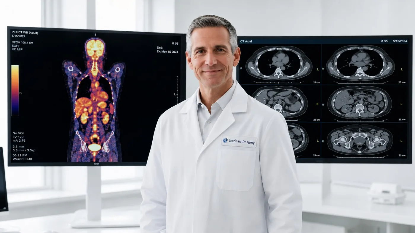
Oncology Imaging Division Highlights

  • Extensive experience in providing services in support of oncology and immuno-oncology clinical trials.
  • 180+ board-certified radiologists focused in Oncology.
  • Participated in hundreds of Phase I through IV Oncology clinical trials for both safety and efficacy.
  • Expertise includes the full spectrum of oncology and immuno-oncology imaging indications and assessments.
  • Experts in all Oncology Criteria: BI-RADS, Cheson, Choi, irRC, iRECIST, IWCLL, Lugano, Macdonald, mRECIST, PCWG-2, PERCIST, RANO, RECIST and others.
  • Modality expertise includes: CT, MRI, PET/CT, SPECT, bone scintigraphy and ultrasound.
  • Integrated CRF with multiple criteria support.

Blinded Independent Central Review (BICR)

To reduce reader variability and bias in oncology trials, our core lab conducts blinded independent central review with adjudication, performed by board-certified radiologists focused in oncology. Readers are trained and qualified on each protocol's imaging charter, and every review follows standardized methodologies with integrated data capture.

Tumor Response Criteria: RECIST 1.1 and Beyond

From RECIST 1.1 solid-tumor assessments to criteria for immunotherapy (iRECIST, irRC), lymphoma (Lugano, Cheson), neuro-oncology (RANO, Macdonald), prostate cancer (PCWG-2), and metabolic response (PERCIST), our integrated case report forms support multiple criteria within a single study.

Frequently Asked Questions

What response criteria does Intrinsic Imaging support for oncology trials?
All major oncology criteria, including RECIST 1.1, iRECIST, mRECIST, irRC, Lugano, Cheson, Choi, RANO, RANO 2.0, Macdonald, PCWG3, PERCIST, IWCLL, BI-RADS and protocol-specific methodologies.
Does Intrinsic Imaging provide blinded independent central review (BICR)?
Yes. Our oncology radiologists perform blinded independent central review and adjudication under standardized, protocol-specific review methodologies.
Which imaging modalities does the oncology core lab support?
CT, MRI, PET/CT, SPECT, bone scintigraphy and ultrasound, including diffusion, perfusion, volumetric and quantitative PET methods across Phase I through IV studies.

Indication-Specific Oncology Imaging and Response Criteria

Our oncology imaging core lab selects the acquisition method, response criteria and independent review design around the tumor type and treatment. The imaging charter defines baseline eligibility, lesion selection, follow-up timing and response rules before central review begins.

Trial settingImaging and criteriaCentral assessment
Solid tumorsCT and MRI; RECIST 1.1Target and non-target lesions, new lesions, best overall response and progression dates
Immuno-oncologyCT and MRI; iRECIST and protocol-defined immune-response criteriaUnconfirmed and confirmed progression, with confirmation timing defined in the protocol
LymphomaFDG PET/CT and CT; Lugano and Deauville five-point scaleMetabolic and anatomic response, nodal and extranodal disease
Neuro-oncologyContrast-enhanced MRI; RANO and RANO 2.0Enhancing and non-enhancing disease, with relevant clinical and corticosteroid information
Prostate cancerCT, MRI and bone scintigraphy; PCWG3Soft-tissue response and bone progression, including protocol-defined confirmation rules
Hepatocellular carcinomaMultiphase CT and MRI; mRECISTViable enhancing tumor rather than total lesion size alone
Gastrointestinal stromal tumorsCT; Choi criteriaChanges in tumor size and attenuation
Metabolic responseFDG PET/CT; PERCISTStandardized uptake measurements with acquisition and reconstruction controls

Quantitative Imaging Biomarkers and Radiomics

We measure lesion dimensions, tumor volume, diffusion-derived apparent diffusion coefficient (ADC), perfusion and PET uptake for efficacy and exploratory biomarker analyses. Radiomics support includes lesion segmentation, extraction of quantitative image features and reproducibility checks across scanners and acquisition settings. Feature definitions and analysis methods are specified before analysis to limit avoidable bias.

Imaging assessments feed endpoints such as objective response rate, duration of response and progression-free survival. The protocol defines how central imaging results are combined with clinical data; imaging alone does not establish overall survival or patient benefit.

Reader Agreement, Adjudication and Longitudinal Consistency

Readers qualify on the study criteria and representative cases before production reads. Independent reviewers follow a defined longitudinal reading sequence, with blinding rules that control access to treatment assignment, site assessments and other information that could bias interpretation.

We track discrepancies in lesion selection, response category and progression timing. An independent adjudicator resolves disagreements according to the charter. Reader agreement and drift reviews identify where calibration or retraining is needed, while preserving an audit trail of original assessments and adjudicated results.

PET/CT and Multicenter Acquisition Controls

Site instructions specify scan coverage, slice thickness, contrast timing and follow-up consistency. PET controls address uptake time, glucose documentation, scanner calibration and reconstruction. Quality control checks whether each examination is suitable for the intended endpoint and records missing scans, non-evaluable images and site queries.

Can one study use more than one response framework?
Yes. We configure separate assessments and data fields for each protocol-defined framework, such as RECIST 1.1 and iRECIST, while preserving independent response rules and traceable results.

Planning an oncology trial? Our subspecialists can help.

Partner With Us

Featured Case Study

Oncology

Phase III Immunotherapy Trial Using RECIST 1.1 and iRECIST

  • 90+ sites in 18 countries
  • CT, MRI
  • RECIST 1.1 and iRECIST

Challenge

The sponsor needed to tell pseudoprogression apart from true disease progression. This had to work consistently across more than 90 sites, using two response frameworks at once.

Our Solution

Subspecialty oncologic radiologists ran dual independent assessments. Automated discordance triggers flagged unconfirmed progression findings and sent them for adjudication.

Results

  • Consistent central assessment of 12500+ timepoints
  • Full audit trail supported the BLA submission
  • Zero Form 483 observations during the sponsor inspection
View all case studies