← All Therapeutic Areas
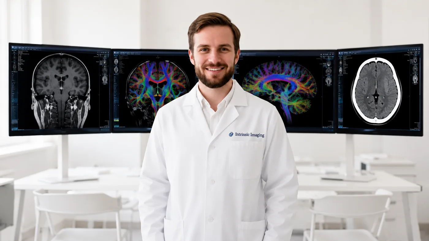
Neuroradiologist in front of brain MRI and DTI tractography monitors

Central Nervous System Imaging Division — Neuroimaging Core Lab

Our Central Nervous System (CNS) Imaging division provides specialized medical imaging expertise for clinical trials evaluating neurological, neurodegenerative, cerebrovascular, psychiatric, and spinal disorders. Supported by a network of more than 100 board-certified and fellowship-trained neuroradiologists, the CNS Imaging division combines deep subspecialty expertise with rigorous, standardized imaging methodologies designed to generate consistent, reproducible, and clinically meaningful data across multicenter and global studies.

Our experience encompasses the full spectrum of brain and spine disease processes, including Alzheimer's disease and other dementias, multiple sclerosis, Parkinson's and Huntington's diseases, epilepsy, stroke and cerebrovascular disease, traumatic brain injury, brain and spinal tumors (with our Oncology Imaging division), spinal cord injury, and degenerative disorders of the spine.

The CNS Imaging division supports sophisticated imaging protocols incorporating structural and functional MRI, MRA, CT and CTA, CT and MR perfusion, diffusion and diffusion tensor imaging, PET and other nuclear imaging techniques, spectroscopy, catheter angiography, and transcranial Doppler ultrasound. Intrinsic Imaging works closely with sponsors from protocol development and imaging charter design through site qualification, image acquisition standardization, quality control, centralized independent review, quantitative analysis, and final data delivery.

Particular emphasis is placed on controlling imaging variability across sites, scanners, readers, and longitudinal timepoints—an essential consideration in CNS trials where subtle changes in anatomy, lesion burden, perfusion, diffusion, metabolism, or other imaging biomarkers may provide critical evidence of disease progression or therapeutic response. Through this combination of subspecialty physician expertise, advanced imaging capabilities, and disciplined central-review methodology, Intrinsic Imaging provides sponsors with the imaging infrastructure and scientific rigor required to support complex CNS clinical development programs.

In CNS trials, imaging biomarkers such as lesion burden, perfusion, diffusion, and metabolism often serve as primary or secondary imaging endpoints. We define each biomarker in the imaging charter and measure it consistently over time, so small changes reflect true disease progression or therapeutic response.

Central Nervous System Imaging Division Highlights

  • Extensive experience in providing services in support of clinical trials involving the central nervous system.
  • 100+ board-certified and fellowship-trained neuroradiologists.
  • Expertise includes the full spectrum of brain and spine disease processes.
  • Modality expertise includes:
    • CT & CTA including CT perfusion
    • Diffusion tensor imaging
    • MRI & MRA including MR perfusion
    • Nuclear imaging including FDG PET, catheter angiography with special expertise in invasive devices
    • Spectroscopy
    • Ultrasound including transcranial doppler

Featured Case Study

Central Nervous System

Phase II Alzheimer's Disease Volumetric MRI and Amyloid PET Study

  • 55 sites
  • 3T MRI, amyloid PET
  • Volumetric and visual PET endpoints

Challenge

Differences between scanner vendors added variability to volumetric MRI measurements and amyloid PET assessments. This spanned 55 academic and commercial imaging centers.

Our Solution

Intrinsic Imaging set up phantom-based site qualification and automatically checked acquisition parameters at submission. Central assessment paired quantitative volumetric analysis of atrophy with standardized visual PET scoring.

Results

  • Cross-site measurement variation reduced by 38%
  • Reliable secondary and exploratory biomarker endpoints
  • Comparable imaging data across scanner vendors
View all case studies

Neuroimaging Endpoints for CNS Clinical Trials

Our neuroimaging core lab supports trials in Alzheimer's disease and other dementias, multiple sclerosis, Parkinson's and Huntington's diseases, epilepsy, stroke, traumatic brain injury, brain and spinal tumors, and spinal cord injury. Endpoints may include lesion burden, structural change, perfusion, diffusion, metabolism, and other imaging biomarkers of disease progression or therapeutic response.

Advanced MRI, PET and Perfusion Imaging

Our neuroradiologists read structural and functional MRI, MRA, CT and CTA, CT and MR perfusion, diffusion and diffusion tensor imaging, spectroscopy, FDG PET and other nuclear imaging, catheter angiography, and transcranial Doppler ultrasound.

Controlling Variability in Multicenter Neuroimaging

Because subtle longitudinal change can be decisive in CNS trials, we emphasize site and scanner qualification, acquisition standardization, centralized quality control, and blinded independent central review with reader training and adjudication.

Frequently Asked Questions

Which neurological indications do you support?
Alzheimer's and variants, multiple sclerosis, Parkinson's, Huntington's, epilepsy, stroke and cerebrovascular disease, brain tumors, traumatic brain injury, spinal disorders, schizophrenia, and more.
Who reads CNS trial images?
More than 100 board-certified, fellowship-trained neuroradiologists.
Do you support perfusion and diffusion imaging?
Yes — CT and MR perfusion, diffusion and diffusion tensor imaging, and spectroscopy are all supported.

Neuroimaging Methods by Disease and Endpoint

CNS trials require methods that distinguish disease progression, treatment effects and imaging safety findings. We align sequences, tracer selection, quantitative analysis and central review with the indication and the role of each endpoint.

IndicationMethodsAssessments
Alzheimer’s disease and dementiaStructural MRI, amyloid PET, tau PET and FDG PETWhole-brain and hippocampal volume, regional tracer uptake and longitudinal change
Anti-amyloid treatment safetyFLAIR and susceptibility-sensitive MRIARIA-E edema or effusion and ARIA-H microhemorrhage or superficial siderosis
Multiple sclerosisT2/FLAIR, contrast-enhanced T1 and volumetric MRINew or enlarging lesions, enhancing lesions and brain-volume change
ALS and motor-neuron diseaseStructural MRI and diffusion tensor imagingRegional atrophy and white-matter measures as protocol-defined exploratory biomarkers
Parkinson’s and movement disordersMRI, dopamine-transporter SPECT and PETRegional structure, tracer binding and protocol-defined functional measures
Stroke and cerebrovascular diseaseCT/CTA, MRI/MRA and perfusion imagingASPECTS, infarct volume, vessel status and perfusion-defined tissue measures
Brain tumorsContrast-enhanced MRI, diffusion and perfusionRANO-based response, enhancing disease and non-enhancing change

ARIA Safety Review and Escalation

For anti-amyloid programs, readers assess amyloid-related imaging abnormalities using the protocol’s severity definitions and comparison timepoints. We distinguish ARIA-E from ARIA-H, document lesion location and extent, and track changes on follow-up MRI. Findings are communicated through an agreed safety-escalation process; treatment decisions remain with the investigator and sponsor.

Volumetric MRI and Molecular Imaging Biomarkers

Quantitative workflows include brain and regional segmentation, hippocampal volumetry, lesion burden, diffusion measures and PET uptake quantification. Registration, segmentation review and acquisition checks help distinguish biological change from motion, scanner differences or analysis artifacts. PET reference regions and any tracer-specific normalization methods are defined in the analysis plan.

Scanner Harmonization and Longitudinal Quality Control

We qualify scanner configurations and standardize sequences, positioning and reconstruction before enrollment. Phantom testing is used where required by the quantitative endpoint. Scanner upgrades, protocol changes and repeat examinations are documented so their effects can be considered in analysis.

Central readers follow prespecified blinding and comparison rules. Image quality, missing sequences and non-evaluable assessments are tracked alongside endpoint results. Calibration cases and adjudication support consistent reads throughout the study.

Are volumetric and diffusion measures always primary endpoints?
No. Their role may be primary, secondary, safety or exploratory, depending on the protocol and supporting validation. We define that role and the analysis method with the sponsor before reads begin.

Direct physician access

Work Directly With Our Medical Leadership

  • • Your medical monitor talks directly to board-certified physicians — no intermediary layers.
  • • Physicians help author your imaging charter and resolve reader discrepancies.
  • • The same leadership team stays with your study from kickoff to database lock.
Meet Our Leadership
  • 3 FDA inspections

    Zero 483 observations

  • 5 BSI ISO certifications

    9001, 13485, 22301, 27001, 27018

  • 1000+ physicians

    Board-certified, fellowship-trained

Planning a central nervous system trial? Our subspecialists can help.

Contact