Intrinsic Imaging
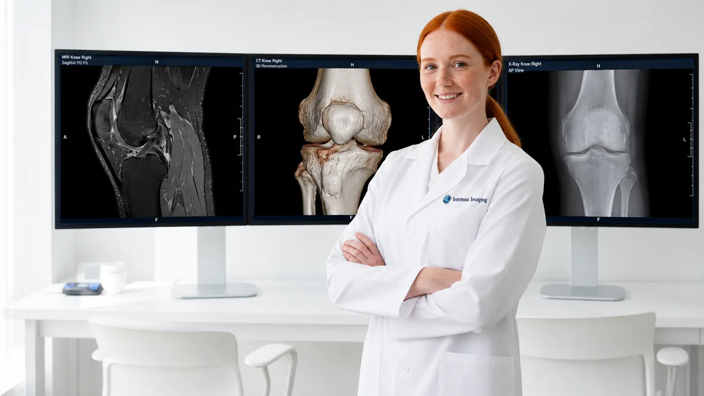
Musculoskeletal radiologist in front of knee MRI, spine X-ray, and skeletal imaging monitors

Musculoskeletal Imaging Core Lab Division

Intrinsic Imaging provides imaging core lab services in support of musculoskeletal clinical trials. Established and study-specific scoring systems, applied by our musculoskeletal radiologists alongside quantitative imaging methods, provide sponsors with objective measures of disease progression and therapeutic response in indications affecting the bones, joints, muscles, connective tissues and spine. Intrinsic Imaging brings this expertise to orthopedic implant and spinal device studies as well.

At Intrinsic Imaging, our Musculoskeletal Imaging division brings together more than 150 board-certified, fellowship-trained musculoskeletal radiologists with expertise across a broad spectrum of musculoskeletal conditions, including osteoarthritis, rheumatoid and inflammatory arthritis, ankylosing spondylitis, osteoporosis, muscular dystrophy, spinal muscular atrophy, and cartilage disorders. This expertise extends to bone healing and repair, orthopedic implants, and other degenerative, inflammatory, metabolic, neuromuscular, and structural diseases affecting the musculoskeletal system.

Intrinsic Imaging supports musculoskeletal endpoints across an extensive range of imaging technologies, including radiography, CT and quantitative CT (QCT), DXA, conventional and advanced MRI, kinetic MRI, metal artifact reduction imaging, nuclear imaging, and ultrasound. Our teams work closely with sponsors from protocol development and imaging charter design through regulatory-ready data delivery to qualify imaging sites, standardize positioning and acquisition, maintain image quality control, train and qualify readers on each scoring system, and conduct centralized independent review with adjudication and quantitative analysis.

By combining musculoskeletal subspecialty expertise with standardized acquisition, calibrated scoring, and reproducible quantitative assessment, Intrinsic Imaging helps sponsors control variability across imaging sites, equipment, readers, and longitudinal timepoints. This consistency provides the methodological rigor needed to detect subtle structural and functional changes in complex musculoskeletal clinical trials.

Musculoskeletal Imaging Division Highlights

  • Extensive experience in providing services for musculoskeletal clinical trials.
  • 150+ board-certified and fellowship-trained musculoskeletal radiologists.
  • Expertise in the full complement joint and spine disease processes.
  • Expertise in all scoring systems for predictable, meaningful data.
  • Modality expertise includes:
    • CT, QCT, DXA
    • X-Ray, MRI & Kinetic MRI
    • MARs imaging, Nuclear imaging
    • Ultrasound

MSK Imaging Endpoints and Scoring Systems

At Intrinsic Imaging, our musculoskeletal imaging core lab implements established and study-specific scoring systems and quantitative methods for endpoints such as joint-space width, cartilage thickness and volume, bone mineral density, structural progression, inflammation, bone healing, implant performance, and spinal motion and kinetics. These methodologies are designed to generate objective, reproducible, and clinically meaningful data across multicenter and global clinical trials.

DXA, Quantitative CT and Advanced MRI

Intrinsic Imaging’s musculoskeletal radiologists centrally read radiography, CT and quantitative CT (QCT), DXA, conventional and kinetic MRI, metal artifact reduction (MARS) imaging, nuclear imaging, and ultrasound for osteoarthritis, rheumatoid and inflammatory arthritis, ankylosing spondylitis, osteoporosis, muscular dystrophy, spinal muscular atrophy, and orthopedic implant trials. Readers are selected for their experience with the indication, the imaging modality, and the scoring method each protocol specifies.

Reader Calibration and Independent Central Review

Through reader training and qualification, centralized quality control, and blinded independent central review with adjudication, Intrinsic Imaging delivers predictable, reproducible MSK data across sites and timepoints.

Musculoskeletal Scoring, Quantification and Medical Device Imaging

Intrinsic Imaging’s MSK core lab combines structural scoring with quantitative imaging for arthritis, bone health, muscle disease, and orthopedic medical device studies. Methods are selected around the tissue, the mechanism of action, and the follow-up interval, aligning each measure with the objectives of the study.

Study focusMethods and scoringMeasures
Rheumatoid arthritisX-ray Sharp/van der Heijde; MRI RAMRISErosions, joint narrowing, synovitis, osteitis, progression
OsteoarthritisMRI MOAKS/WORMS; X-ray Kellgren-LawrenceCartilage, marrow lesions, menisci, joint-space width
Bone healthDXA, QCTAreal/volumetric BMD; site and scanner calibration
Cartilage repairMorphologic and quantitative MRIThickness, volume, defect filling, composition
Muscle diseaseMRI; Dixon fat quantificationMuscle volume, fat fraction, tissue change
Implants and fracture healingX-ray, CT, metal artifact reduction MRIAlignment, fusion, healing, implant and peri-implant findings

Arthritis Reader Calibration and Scoring

Before evaluating study images, Intrinsic Imaging’s readers train on the selected atlas and scoring conventions. The imaging charter defines which joints or compartments are scored, how longitudinal pairs are presented, and how missing anatomy is handled, while adjudication and agreement monitoring address differences that could affect structural-progression endpoints.

Bone Density and Quantitative Cartilage Imaging

Intrinsic Imaging’s DXA workflows define anatomical regions, positioning, analysis boundaries, and cross-calibration requirements. QCT and quantitative MRI require consistent acquisition and reconstruction, and phantom checks are included where the endpoint depends on them. Segmentation and measurement quality are reviewed before results are released.

Intrinsic Imaging analyzes cartilage and muscle measures using prespecified methods, with thresholds fixed before results are known. Scanner changes, motion, and metal artifacts are documented as potential sources of variability.

Emerging Indication Spotlight: Rare Neuromuscular Disease and Muscle MRI

Gene therapies and other targeted treatments for rare neuromuscular conditions, including Duchenne and other muscular dystrophies, spinal muscular atrophy, and inflammatory myopathies, increasingly rely on quantitative muscle MRI. Small patient populations make every assessment valuable, and Intrinsic Imaging supports these trials with imaging biomarkers that can detect change sooner than functional tests and strengthen a study design.

Intrinsic Imaging applies Dixon-based fat-fraction quantification, muscle volumetry, and T2 mapping to protocol-specified muscle groups, with standardized positioning and coverage across visits. Segmentation follows documented anatomical rules and is reviewed for consistency, and sites in a dispersed rare-disease network are qualified with test submissions before enrollment. Results are delivered with evaluability flags for integration with functional and clinical outcome measures.

Orthopedic Device Follow-Up

Intrinsic Imaging standardizes imaging views and anatomical landmarks for implants, spinal devices, and fracture-healing studies, allowing readers to assess positioning, structural change, and relevant complications consistently over time. Image-derived findings are delivered with quality and evaluability flags for integration with the sponsor’s clinical assessments.

Guidance for Sponsors

Physician-authored guidance from Intrinsic Imaging on imaging endpoints and trial design.

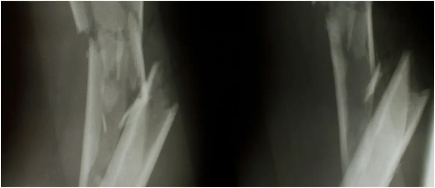
Radiographs of a comminuted long-bone diaphyseal fracture

Guidance for Sponsors · Volume 4, Issue 4 · 2026 Edition

Imaging Analysis and Operative Treatment of Diaphyseal Fractures

Fractures are among the most common reasons for an imaging examination, and imaging guides every stage of care: diagnosis, classification, reduction, fixation, detection of complications and confirmation of healing. Radiography, CT, MRI and bone scintigraphy each play a distinct role. For sponsors of orthopedic drug, biologic and device trials, the central challenge is that fracture union, the most commonly reported primary outcome, still has no universally accepted radiographic definition. Standardized scoring, consistent imaging schedules and independent central review turn a subjective judgment into a defensible endpoint.

Read the article
Illustration of the human spine and skeleton

Guidance for Sponsors · Volume 4, Issue 5 · 2026 Edition

Orthopedic Device Assessments: Medical Imaging Challenges in Clinical Trials

Orthopedic research spans joint reconstruction, spinal fusion and motion preservation, and biologics for cartilage, tendon and ligament repair. Regulators and payers expect rigorous quantitative and qualitative evidence of efficacy, safety and longevity, and imaging supplies much of it. Since investigational devices are often unlike anything imaged before, protocols must be built around the device itself: its engineering, materials, surgical technique and position. Advances in CT, metal-artifact-reduction MRI and radiostereometric analysis now allow earlier and more precise answers than ever.

Read the article
Illustration of an inflamed knee joint

Guidance for Sponsors · Volume 4, Issue 8 · 2026 Edition

Advances in High-Field MRI for the Assessment of Arthritis

Arthritis is a leading cause of disability, affecting about 53 million U.S. adults. Its central feature is loss of hyaline cartilage, which has little capacity to repair. Radiographs show only late damage; high-field 3.0 Tesla MRI shows cartilage morphology in fine detail, and compositional techniques such as T2 mapping reveal degeneration before visible loss. For sponsors, the challenge is regulatory as well as technical: FDA has not yet accepted a structural endpoint for osteoarthritis approval, so imaging must be precise, reproducible and linked to how patients feel and function.

Read the article

More guidance: Osteoporosis Imaging in Trials; Knee Osteoarthritis Imaging in Trials; Quantitative MRI in Orthopedic Trials; Spinal Deformity Imaging in Trials; Rheumatoid Arthritis Imaging in Trials

Browse all Guidance for Sponsors →

Featured Case Study

Intrinsic Imaging

Musculoskeletal

Phase III Knee Osteoarthritis DMOAD Trial

  • 24-month follow-up
  • Knee MRI, radiographs
  • JSN and WORMS

Challenge

Evaluating disease modification in knee osteoarthritis requires reproducible measurement of submillimeter changes in joint space narrowing over extended follow-up. The sponsor required readings of sufficient precision to register these minute structural changes, together with consistent tracking of WORMS scores across 24 months of serial MRI examinations. Since the primary structural endpoint rested on longitudinal comparison, any drift in reader interpretation or measurement technique over the study's duration would directly compromise the trial's conclusions.

Read the case study
View all case studies

Frequently Asked Questions

Is Intrinsic Imaging independently owned?
Yes. Intrinsic Imaging returned to independent, management-led ownership in 2026 after five years within WCG Clinical. Todd A. Joron and Dr. Amit Mehta reacquired the company. Intrinsic Imaging’s CEO letter describes the return to independence as restoring agility, direct accountability and strategic focus.
What is Intrinsic Imaging’s FDA Establishment Identifier?
Intrinsic Imaging’s current FDA Establishment Identifier (FEI) is 3013568226. The August 2012 inspection was recorded under FEI 3009573796 at the company’s former San Antonio, Texas address.
How long does Intrinsic Imaging typically take for charter and reading system setup?
Intrinsic Imaging typically finalizes the imaging charter within about 20 business days of kickoff and prepares the reading system within about 15 business days. Intrinsic Imaging agrees the dependencies and sequence for these distinct startup milestones with the sponsor for each study.
Go to more Frequently Asked Questions

Tell us about your trial.

Planning a Musculoskeletal trial? Our team will respond within one business day.