
Guidance for Sponsors · Volume 5, Issue 11 · 2026 Edition
Quantitative MRI Imaging in Orthopedic Clinical Trials
By Jennifer Swart, M.D., Board Certified Radiologist · Updated for 2026 by Intrinsic Imaging
Download the PDF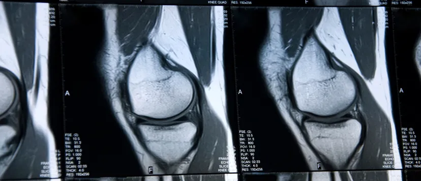
Orthopedic trials have long depended on patient questionnaires and on radiographic joint space width, a measurement of what cannot be seen. Quantitative MRI measures cartilage, bone and soft tissue directly, in three dimensions, and can reveal biochemical change before structural loss occurs. Validated scoring systems for rheumatoid arthritis, consensus profiles for compositional cartilage imaging and metal artifact reduction techniques for implants have made MRI a practical endpoint across arthritis and device trials. This issue reviews those methods and what sponsors must standardize to use them well.
Imaging-Related Endpoints
Historically, trials of therapies for arthritis relied primarily on patient questionnaires that assess pain, quality of life and joint function. These measures are inherently subjective and require that a patient exhibit overt symptoms, which limits their value for interventions that aim to slow or halt cartilage loss before symptoms emerge.
Orthopedic efficacy trials therefore combine clinical, laboratory and imaging outcomes, and imaging measurements are often primary endpoints. This is especially true in rheumatoid arthritis and osteoarthritis, and in patients who have received spinal fusion implants, fracture fixation devices or joint reconstruction hardware. The imaging core laboratory provides independent, semiquantitative or fully quantitative analysis of the images to assess the efficacy of the intervention.
The Limits of Radiography
In osteoarthritis and rheumatoid arthritis, structural damage to articular cartilage is a principal measure of progression, traditionally assessed with serial radiographs of joint space width and joint space narrowing. These measurements are reasonably objective but insensitive: trials typically require large samples followed for several years.[1] They are also measurements of negative space, the gap between adjacent bones, and that gap reflects not only cartilage but also subchondral bone deformity and meniscal loss.
Quantitative MRI
Unlike arthroscopy, which is invasive and evaluates only the cartilage surface, and radiography, which infers cartilage from a two-dimensional gap, MRI visualizes the full thickness and volume of cartilage. Three-dimensional segmentation of high-resolution sequences yields cartilage thickness, volume and surface area for each region of the joint. These quantitative morphometric measures are more sensitive to change than radiography and can detect treatment effects in smaller trials over shorter periods.
Compositional Imaging
Cartilage composition changes before cartilage is lost. T2 mapping reflects collagen network integrity and water content, and T1ρ is sensitive to proteoglycan depletion. The 2021 QIBA profile for compositional imaging of knee cartilage set out the hardware, acquisition and analysis practices needed for reliable, reproducible measurement across sites.[2] Delayed gadolinium-enhanced MRI of cartilage (dGEMRIC) also measures proteoglycan content, but it requires a gadolinium-based contrast agent and a prolonged delay before imaging. Since FDA's 2017 class warning on gadolinium retention in the body,[3] noncontrast techniques have generally been preferred for repeated measurement in trials.
Rheumatoid Arthritis
In rheumatoid arthritis, MRI detects synovitis, bone marrow edema (osteitis) and erosions earlier and more sensitively than radiography, and bone marrow edema predicts subsequent erosive progression. The OMERACT Rheumatoid Arthritis MRI Scoring System (RAMRIS) is the validated standard for the hands and wrists, and its 2017 update incorporated additional features, including tenosynovitis and joint space narrowing.[4] RAMRIS allows trials to demonstrate structural benefit with fewer patients and shorter follow-up than radiographic scoring.
Orthopedic Implants
MRI is increasingly used to evaluate joint replacements and fixation hardware, detecting periprosthetic osteolysis, infection, loosening and the soft-tissue reactions associated with metal wear debris. Metal causes susceptibility artifact that distorts images, but metal artifact reduction sequences (MARS), multiacquisition variable-resonance image combination (MAVRIC) and slice-encoding metal artifact correction (SEMAC) have greatly reduced it.[5] These techniques depend on scanner hardware and software, and their availability varies across sites. MR safety and conditional labeling of each device must also be confirmed before imaging.
Guidance for Sponsors
Using quantitative MRI in an orthopedic trial
- Agree on endpoints early. Discuss MRI-based structural endpoints with FDA before the pivotal trial.[1]
- Qualify every site. Verify scanner capability, sequences and phantom performance before enrollment.[2]
- Prefer noncontrast techniques. Use T2 or T1ρ mapping rather than dGEMRIC for repeated measurement.[2,3]
- Use validated scoring. Apply RAMRIS in rheumatoid arthritis with calibrated central readers.[4]
- Plan for metal. Specify artifact reduction sequences and confirm MR conditional status for implant trials.[5]
- Read centrally. Use blinded, independent analysis under a documented imaging charter.[6]
Summary
Quantitative MRI has matured from a research technique into a practical trial endpoint. It measures cartilage directly, reveals composition before structure fails, scores inflammatory disease with validated systems and, with artifact reduction, evaluates the tissue around implants. Sponsors who standardize acquisition across sites and rely on expert central analysis gain endpoints that are more sensitive, more efficient and more informative than radiography alone.
About the Author
Dr. Jennifer Swart, a board-certified radiologist with specialized training in orthopedic radiology, wrote the original edition of this issue at Intrinsic Imaging. She graduated summa cum laude in cognitive neuroscience from Harvard University and earned her M.D. from Columbia University College of Physicians and Surgeons, where she received the Janeway Prize, awarded to the graduate at the top of the class. As an American Academy of Neurology research scholar, she conducted research in neurology published in the journal Stroke. She completed her diagnostic radiology residency at Johns Hopkins Hospital in Baltimore, where she was chief resident, and subspecialized in musculoskeletal radiology at National Orthopedic Imaging Associates in San Francisco with Dr. David Stoller.
References
- U.S. FDA. Osteoarthritis: structural endpoints for the development of drugs, devices, and biological products for treatment. Draft guidance, August 2018. https://www.fda.gov/regulatory-information/search-fda-guidance-documents/osteoarthritis-structural-endpoints-development-drugs
- Chalian M, Li X, Guermazi A, et al. The QIBA profile for MRI-based compositional imaging of knee cartilage. Radiology. 2021;301(2):423–432. https://pubmed.ncbi.nlm.nih.gov/34491127/
- U.S. FDA. Drug safety communication: FDA warns that gadolinium-based contrast agents (GBCAs) are retained in the body; requires new class warnings. December 2017. https://www.fda.gov/media/111506/download
- Østergaard M, Peterfy CG, Bird P, et al. The OMERACT Rheumatoid Arthritis Magnetic Resonance Imaging (MRI) Scoring System: updated recommendations by the OMERACT MRI in Arthritis Working Group. J Rheumatol. 2017;44(11):1706–1712. https://pubmed.ncbi.nlm.nih.gov/28811353/
- Feuerriegel GC, Sutter R. Managing hardware-related metal artifacts in MRI: current and evolving techniques. Skeletal Radiol. 2024;53(9):1737–1750. https://pmc.ncbi.nlm.nih.gov/articles/PMC11303499/
- U.S. FDA. Clinical trial imaging endpoint process standards: guidance for industry. April 2018. https://www.fda.gov/regulatory-information/search-fda-guidance-documents/clinical-trial-imaging-endpoint-process-standards-guidance-industry
Planning an orthopedic or arthritis trial?
Talk with our team about quantitative MRI protocols, RAMRIS and cartilage analysis, and independent central review.
Contact UsRelated reading: Advances in High-Field MRI for the Assessment of Arthritis · Medical Imaging of Osteoarthritis of the Knee in Clinical Trials · Orthopedic Device Assessments: Medical Imaging Challenges in Clinical Trials