← All Therapeutic Areas
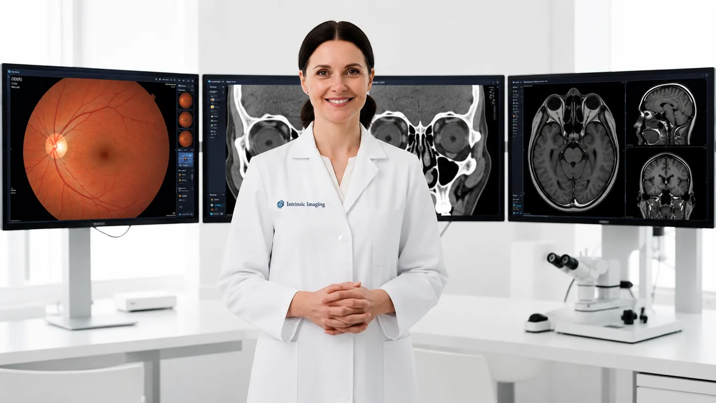
Ophthalmologist in front of OCT retinal scans and fundus photography monitors

Ophthalmology Imaging Division — Ophthalmic Imaging Reading Center

Our Ophthalmology Imaging division provides specialized imaging, functional assessment, and centralized independent review expertise for clinical trials evaluating retinal, macular, optic nerve, and other ophthalmic diseases. Our board-certified ophthalmologists bring extensive clinical research experience, having participated in more than 160 ophthalmology clinical trials as principal investigators or expert reviewers.

The Ophthalmology Imaging division supports studies across a broad spectrum of ophthalmic indications, including diabetic retinopathy, age-related macular degeneration, retinitis pigmentosa and other inherited retinal diseases, epiretinal membrane, macular hole, retinal tears and detachment, and other conditions in which precise characterization of ocular structure and visual function is critical to evaluating disease progression and therapeutic response.

The Ophthalmology Imaging division provides expertise across an extensive range of structural, functional, angiographic, and electrophysiologic assessments, including optical coherence tomography (OCT), color fundus photography (CFP), fundus autofluorescence (FAF), near-infrared autofluorescence, fluorescein angiography, adaptive optics retinal imaging, best-corrected visual acuity (BCVA), visual field assessments, electroretinography (ERG), full-field stimulus testing (FST), intraocular pressure (IOP), and functional MRI. Intrinsic Imaging works closely with sponsors to develop study-specific ophthalmic imaging and assessment strategies encompassing protocol development, imaging charter design, site and equipment qualification, acquisition standardization, image quality control, reader training and qualification, centralized independent review, adjudication, quantitative analysis, and regulatory-ready data delivery.

Particular emphasis is placed on minimizing variability across sites, imaging systems, operators, readers, and longitudinal visits—providing the consistency and methodological rigor necessary to detect subtle anatomical and functional changes over time. By integrating ophthalmic subspecialty expertise with standardized image acquisition, sophisticated quantitative assessment, and disciplined central-review methodologies, Intrinsic Imaging provides the scientific and operational infrastructure required to support complex ophthalmology clinical development programs from early-phase investigation through pivotal trials.

Ophthalmology Imaging Division Highlights

  • Extensive experience delivering specialized services in support of ophthalmic clinical trials across multiple therapeutic areas.
  • Board-certified Ophthalmologists.
  • Participated in 160+ ophthalmology clinical trials as principal investigators or reviewers.
  • Expertise includes full spectrum of ophthalmology imaging and assessments.
  • Modality expertise includes:
    • Optical Coherence Tomography (OCT)
    • Color Fundus Photography (CFP)
    • Adaptive Optics (AO) retinal imaging
    • Best Corrected Visual Acuity (BCVA)
    • Electroretinography (ERG)
    • Fluorescein Angiography
    • Full Field Stimulus Testing (FST)
    • Functional MRI
    • Fundus Autofluorescence (FAF)
    • Intra Ocular Pressure (IOP)
    • Near infrared Autofluorescence (AF) imaging
    • Visual Field Assessments

Featured Case Study

Ophthalmology

Phase II Geographic Atrophy and Neovascular AMD Trial

  • SD-OCT, fundus autofluorescence
  • Lesion area measurement
  • Multi-reader consensus

Challenge

The trial needed precise central measurement of geographic atrophy area. It also needed consistent tracking of choroidal neovascularization over time.

Our Solution

The ophthalmic reading team segmented lesions and used multi-reader consensus to settle unclear lesion boundaries.

Results

  • Lesion area tracking accurate to within 0.05 mm²
  • Primary endpoint readout delivered on schedule
  • Fully audited longitudinal measurements
View all case studies

Ophthalmic Reading Center Services

Our ophthalmic imaging reading center provides centralized independent review of structural, functional, angiographic, and electrophysiologic assessments for trials in diabetic retinopathy, age-related macular degeneration, retinitis pigmentosa and other inherited retinal diseases, epiretinal membrane, macular hole, and retinal tears and detachment.

OCT, Fundus Imaging and Visual Function Endpoints

We support optical coherence tomography (OCT), color fundus photography, fundus and near-infrared autofluorescence, fluorescein angiography, adaptive optics retinal imaging, best-corrected visual acuity, visual fields, ERG, full-field stimulus testing, intraocular pressure, and functional MRI.

Site and Equipment Qualification

Site and equipment qualification, acquisition standardization, image quality control, and reader training keep ophthalmic data consistent across devices, sites, and timepoints.

Frequently Asked Questions

What ophthalmology trial experience do your specialists have?
Our board-certified ophthalmologists have participated in more than 160 ophthalmology clinical trials as principal investigators or reviewers.
Do you read OCT and fundus autofluorescence?
Yes, along with color fundus photography, fluorescein angiography, and adaptive optics imaging.
Do you support functional endpoints?
Yes — BCVA, visual fields, ERG, full-field stimulus testing, and IOP.

Retinal Imaging, Grading and Visual Function Assessments

Our ophthalmic reading center supports structural, angiographic and functional endpoints with equipment-specific acquisition instructions and centralized quality control. We distinguish image-derived outcomes from visual-function testing while coordinating both within the study.

Indication or endpointMethodsMeasures
Neovascular AMD and diabetic macular edemaOCT and fluorescein angiographyCentral subfield thickness, retinal fluid, lesion features and leakage
Geographic atrophyFundus autofluorescence, color photography and OCTAtrophy area, lesion growth and outer-retinal structural change
Diabetic retinopathyStandardized color fundus photographyETDRS diabetic retinopathy severity grading and change over time
Inherited retinal diseaseOCT, autofluorescence and electrophysiologyRetinal structure, ellipsoid-zone measures and protocol-defined functional response
GlaucomaOCT, optic-disc imaging and perimetryRetinal nerve fiber layer, ganglion-cell measures and visual-field change
Visual functionBCVA, visual fields, ERG and full-field stimulus testingSeparate standardized functional assessments matched to the indication

OCT Segmentation and Retinal Fluid Review

OCT quality control checks signal, scan centering, coverage and segmentation. Readers review automated boundaries and apply documented correction rules before measurements are released. Fluid compartments, retinal thickness and anatomical changes are assessed consistently across visits, with device and software changes recorded.

Fundus Grading and Angiographic Endpoints

Fundus photography protocols specify field coverage, illumination and image quality. Graders use the selected disease-severity scale and comparison rules. Fluorescein angiography review addresses phase timing, leakage and lesion characterization; autofluorescence supports quantitative atrophy-area assessments with traceable boundaries.

Site, Photographer and Equipment Qualification

We qualify imaging equipment and personnel through acquisition instructions and representative submissions. Site feedback addresses focus, centering, field coverage and incomplete examinations. Repeat training and quality monitoring help keep acquisition consistent across devices and follow-up visits.

Image grading and functional testing have separate training and data checks. Their results are delivered with evaluability flags and documented query resolution so sponsors can integrate structural and functional endpoints without treating them as equivalent.

Direct physician access

Work Directly With Our Medical Leadership

  • • Your medical monitor talks directly to board-certified physicians — no intermediary layers.
  • • Physicians help author your imaging charter and resolve reader discrepancies.
  • • The same leadership team stays with your study from kickoff to database lock.
Meet Our Leadership
  • 3 FDA inspections

    Zero 483 observations

  • 5 BSI ISO certifications

    9001, 13485, 22301, 27001, 27018

  • 1000+ physicians

    Board-certified, fellowship-trained

Planning an ophthalmology trial? Our subspecialists can help.

Partner With Us