← All Therapeutic Areas
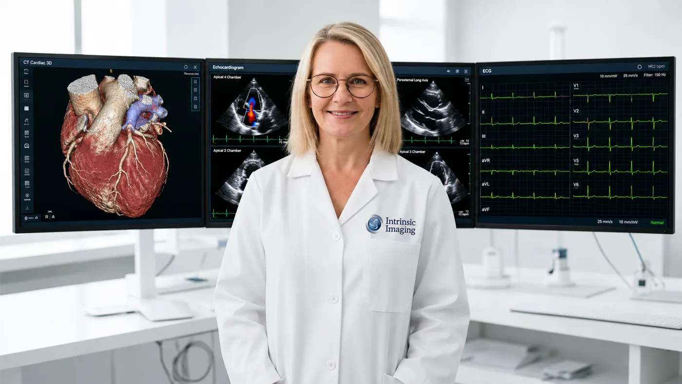
Cardiologist in front of cardiac MRI and coronary CT angiography monitors

Cardiology Imaging Division — Cardiac Imaging Core Lab

Intrinsic Imaging provides comprehensive cardiovascular imaging core lab services for pharmaceutical, biotechnology, and medical device clinical trials, combining subspecialty physician expertise, advanced quantitative imaging, and rigorous image management methodologies to support the evaluation of cardiovascular safety and efficacy.

Our Cardiology Imaging division brings together more than 150 board-certified, fellowship-trained cardiologists and cardiothoracic radiologists with expertise across a broad spectrum of cardiovascular and cardiopulmonary diseases, including heart failure, coronary artery disease, acute coronary syndromes, myocardial infarction, left ventricular dysfunction, valvular and structural heart disease, pulmonary arterial hypertension, peripheral vascular disease, and venous thromboembolic disease.

Intrinsic Imaging supports cardiovascular endpoints across an extensive range of imaging and diagnostic modalities, including 2D and 3D echocardiography, cardiac CT and CT angiography, cardiac MRI and MRA, adenosine stress MRI, invasive angiography, ECG, myocardial strain and speckle-tracking imaging, MUGA, PET, PET/CT, SPECT, nuclear cardiology, vascular ultrasound, and advanced thoracic imaging. Our teams work closely with sponsors from protocol development through database lock to establish standardized acquisition requirements, qualify and train imaging sites, define quantitative and qualitative assessment methodologies, conduct blinded independent central reviews, and implement adjudication procedures designed to minimize inter- and intra-reader variability.

By combining cardiovascular subspecialty expertise with standardized imaging protocols, centralized quality control, and reproducible quantitative assessment, Intrinsic Imaging helps sponsors generate reliable, scientifically robust imaging data suitable for longitudinal endpoint evaluation, clinical development decision-making, and global regulatory submissions.

Cardiology Imaging Division Highlights

  • Extensive experience in providing services in support of clinical trials evaluating cardiac safety and efficacy.
  • 150+ board-certified, fellowship-trained cardiologists and cardiothoracic radiologists.
  • Extensive experience in cardiothoracic imaging indications.
  • Focus on reducing intra-inter cardiothoracic reviewer variability.
  • Modality expertise includes:
    • 2D and 3D Echocardiography
    • Angiography, Cardiac CTA, Cardiac MRI, Chest X-Ray
    • CT, QCT, PET/CT, CR/DR, DXA
    • ECG, MRA, MRI, MRE, Adenosine Stress MRI, MUGA
    • Myocardial Strain imaging, Nuclear imaging
    • PET, SPECT, Speckle tracking, Thoracic CT/MRI, HRCT
    • Ultrasound, Vascular CTA, MRI

Featured Case Study

Cardiology

Global Cardiovascular Outcomes Trial With CCTA and Cardiac MRI

  • 60 sites
  • CCTA, cardiac MRI
  • Plaque, stenosis and LVEF

Challenge

The trial needed reproducible plaque measurement, stenosis grading and ejection fraction readings. This had to work across 60 sites using different scanner models.

Our Solution

Intrinsic Imaging set standard acquisition guidelines. We added a dedicated quality control gateway to catch unusable scans before they reached central review.

Results

  • 99.1% evaluable scan rate across 3400+ patients
  • Inter-reader variability maintained below 4%
  • Ongoing reader calibration throughout the trial
View all case studies

Cardiac Safety and Efficacy Imaging Endpoints

Our cardiac imaging core lab supports clinical trials evaluating both cardiac safety and cardiovascular efficacy. Endpoints are defined with sponsors during protocol and imaging charter development, then assessed by board-certified, fellowship-trained cardiologists and cardiothoracic radiologists using standardized quantitative and qualitative methods.

Typical assessments include left ventricular function and dysfunction, myocardial strain and speckle tracking, coronary and vascular evaluation by angiography and CTA, valvular and structural heart disease, and pulmonary and thoracic findings across heart failure, coronary artery disease, pulmonary arterial hypertension, and venous thromboembolic disease trials.

Echocardiography, Cardiac MRI and Cardiac CT Core Lab

We centrally read 2D and 3D echocardiography, cardiac MRI and MRA, adenosine stress MRI, cardiac CTA, invasive angiography, ECG, MUGA, PET, SPECT, and nuclear cardiology. Site qualification, acquisition standardization, and centralized quality control keep data consistent across sites, scanners, and longitudinal timepoints.

Blinded Independent Central Review and Adjudication

Blinded independent central review (BICR) is performed under single, double, or triple-read paradigms with adjudication procedures designed to minimize inter- and intra-reader variability, supporting reliable endpoint evaluation and global regulatory submissions.

Frequently Asked Questions

Which cardiac imaging modalities does your core lab read?
Echocardiography (2D/3D), cardiac MRI and MRA, stress MRI, cardiac and vascular CTA, angiography, ECG, MUGA, PET, SPECT, nuclear cardiology, ultrasound, and thoracic CT/MRI/HRCT.
Do you support cardiac safety as well as efficacy endpoints?
Yes. The Cardiology Imaging division supports clinical trials evaluating both cardiac safety and cardiovascular efficacy.
Who performs the reads?
More than 150 board-certified, fellowship-trained cardiologists and cardiothoracic radiologists.

Cardiovascular Imaging Measures and Acquisition Standards

We translate cardiac and vascular trial objectives into reproducible measurements of function, structure and treatment response. Each modality has its own acquisition instructions, measurement conventions and quality checks.

MethodMeasuresTrial applications
2D and 3D echocardiographyLVEF, chamber volumes, global longitudinal strain and Doppler measuresHeart failure, cardiotoxicity, valvular and structural heart studies
Cardiac MRIVentricular volumes, ejection fraction, myocardial mass, late gadolinium enhancement and T1/T2 mappingCardiomyopathy, fibrosis, inflammation and treatment response
Cardiac and vascular CT angiographyStenosis, plaque burden, vessel dimensions and device geometryCoronary disease, vascular interventions and structural devices
Angiography, IVUS and OCTLumen dimensions, lesion characteristics and stent appositionCoronary interventions and device performance
PET, SPECT and MUGAPerfusion, viability and ventricular functionIschemia, myocardial viability and cardiac safety

Echocardiography and Cardiac MRI Measurement Workflows

Echo instructions define required views, frame rates, Doppler recordings and the cardiac cycles used for analysis. Readers apply consistent endocardial tracing and strain methods, with documented rules for poor acoustic windows or incomplete acquisitions.

Cardiac MRI protocols define cine coverage, temporal resolution, tissue-characterization sequences and contrast timing. Contouring conventions address papillary muscles, basal slices and ventricular boundaries to support consistent volumes and function across longitudinal visits.

Vascular and Structural Device Assessments

Central review measures anatomy, lesion severity and device-related findings using protocol-defined landmarks. Quantitative angiography and intravascular imaging support lumen and vessel analysis, while CT and echo assess structural-device positioning and functional effects. Imaging findings are kept distinct from clinical event adjudication unless both are included in the study scope.

Reproducibility Across Equipment and Readers

Site qualification addresses scanner or ultrasound platform, software version and acquisition settings. We document equipment changes, assess image suitability, calibrate readers on measurement conventions and use adjudication for endpoint-relevant disagreements. The data package includes measurement results, quality flags and traceable review records.

Direct physician access

Work Directly With Our Medical Leadership

  • • Your medical monitor talks directly to board-certified physicians — no intermediary layers.
  • • Physicians help author your imaging charter and resolve reader discrepancies.
  • • The same leadership team stays with your study from kickoff to database lock.
Meet Our Leadership
  • 3 FDA inspections

    Zero 483 observations

  • 5 BSI ISO certifications

    9001, 13485, 22301, 27001, 27018

  • 1000+ physicians

    Board-certified, fellowship-trained

Planning a cardiology trial? Our subspecialists can help.

Contact