← All Therapeutic Areas
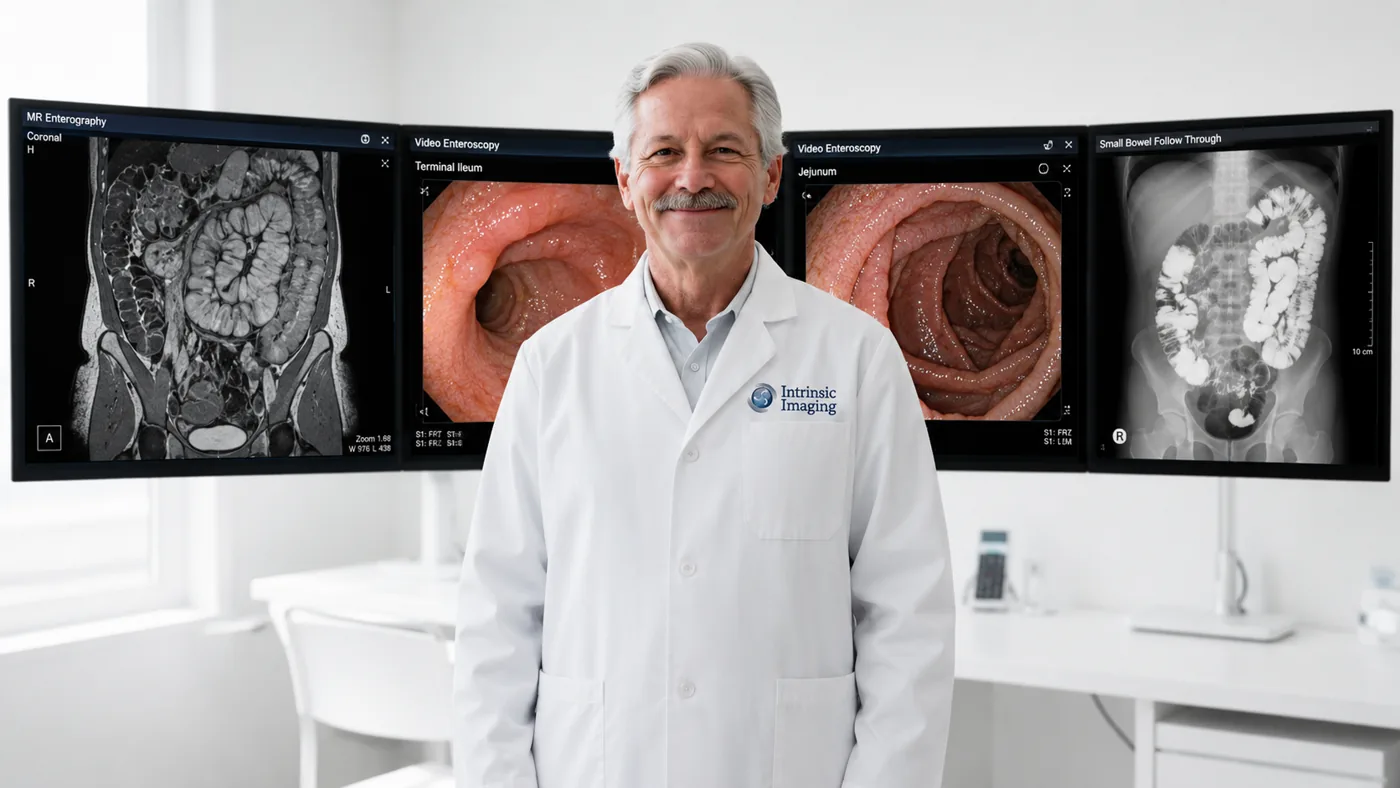
Gastroenterologist in front of abdominal CT, MR enterography, and endoscopy monitors

Gastroenterology Imaging Division — GI Imaging & Endoscopy Core Lab

Our Gastroenterology Imaging division provides specialized imaging and independent review expertise for clinical trials evaluating gastrointestinal, hepatobiliary, pancreatic, metabolic, and related abdominal and pelvic disorders. Supported by a network of more than 60 board-certified and fellowship-trained gastroenterologists, the Gastroenterology Imaging division combines deep clinical subspecialty expertise with rigorous imaging and assessment methodologies designed to produce consistent, reproducible, and clinically meaningful data across multicenter and global clinical trials.

Our experience spans a broad range of acute and chronic gastrointestinal conditions, including inflammatory bowel disease, ulcerative colitis, gastroesophageal reflux disease, short bowel syndrome, gastrointestinal ulcerative disease, and other complex disorders in which imaging, endoscopic evaluation, and objective assessment may play an important role in evaluating disease activity, progression, and therapeutic response.

The Gastroenterology Imaging division supports a comprehensive range of imaging and diagnostic modalities, including CT and high-resolution CT, low-dose and advanced CT techniques, MRI, endoscopy, and nuclear imaging with quantitative methodologies. Intrinsic Imaging works collaboratively with sponsors to develop study-specific imaging strategies encompassing protocol development, imaging charter design, site and reader qualification, acquisition standardization, image and endoscopy quality control, centralized independent review, quantitative assessment, adjudication, and regulatory-ready data delivery.

Particular attention is given to minimizing inter-site and inter-reader variability and maintaining consistent assessment methodologies across longitudinal timepoints—critical considerations when imaging or endoscopic findings contribute to efficacy, safety, disease-activity, or exploratory biomarker endpoints. By integrating experienced gastroenterology specialists with disciplined central-review processes and sophisticated imaging capabilities, Intrinsic Imaging provides the scientific, clinical, and operational expertise required to support complex gastroenterology clinical development programs from early-phase investigation through pivotal trials.

Gastroenterology Imaging Division Highlights

  • Extensive experience in providing services in support of clinical trials involving the GI system.
  • 60+ board-certified and fellowship-trained gastroenterologists.
  • Experience in imaging of organ specific endocrine disorders as well as multiple endocrine syndromes and neoplasia.
  • Modality expertise includes:
    • CT including novel techniques
    • Endoscopy
  • High Resolution CT imaging and low dose techniques.
    • MRI
    • Nuclear imaging with quantitative techniques

Featured Case Study

Gastroenterology

Phase IIb Inflammatory Bowel Disease Trial

  • Crohn's disease and ulcerative colitis
  • Endoscopy video, MR enterography
  • SES-CD and Mayo Endoscopic Score

Challenge

Site-level reading of endoscopic video and MR enterography brought a lot of subjectivity into disease activity assessments.

Our Solution

Intrinsic Imaging set up a blinded dual-reader central review process. It used anonymized video, standardized ulcer measurement and quantitative review of bowel wall thickness.

Results

  • Inter-reader scoring discordance reduced by 42%
  • Reduced incidence of inappropriate screening exclusions
  • Consistent eligibility decisions across sites
View all case studies

Central Endoscopy and GI Imaging Review

Our GI imaging and endoscopy core lab provides centralized independent review of endoscopy and imaging for inflammatory bowel disease, ulcerative colitis, GERD, short bowel syndrome, ulcerative disease, and other gastrointestinal and abdominal disorders where objective assessment of disease activity matters.

CT, MRI and Quantitative Nuclear Imaging

We support high-resolution and low-dose CT, advanced CT techniques, MRI, endoscopy, and nuclear imaging with quantitative methodologies, with study-specific acquisition standards and image and endoscopy quality control.

Consistent Assessment Across Sites and Timepoints

Site and reader qualification, adjudication, and consistent methodology across longitudinal timepoints minimize inter-site and inter-reader variability for efficacy, safety, disease-activity, and exploratory biomarker endpoints.

Frequently Asked Questions

Do you provide central reading of endoscopy?
Yes. Endoscopy is centrally reviewed alongside CT, MRI, and nuclear imaging.
Which GI indications do you support?
IBD, ulcerative colitis, GERD, IBS, short bowel, ulcers, and related abdominal and pelvic conditions.
Who reviews GI trial data?
More than 60 board-certified, fellowship-trained gastroenterologists.

IBD, Liver Disease and Quantitative Abdominal Imaging

GI studies often combine endoscopy, cross-sectional imaging and tissue assessment. We define each assessment separately and coordinate their acquisition, independent review and data delivery around the protocol.

Study focusMethodsCentral assessment
Crohn’s diseaseIleocolonoscopy and MR enterographySES-CD, CDEIS and MaRIA or simplified MaRIA, as specified by the protocol
Ulcerative colitisEndoscopyMayo endoscopic subscore and UCEIS
Steatotic liver disease and MASHMRI-PDFF and MR elastographyLiver fat fraction and stiffness, with acquisition and analysis controls
Liver fibrosisMR or ultrasound elastographyProtocol-defined stiffness measurements and longitudinal change
GI tissue endpointsDigital pathology and histopathology reviewProtocol-defined tissue scores, including Robarts or Nancy indices where applicable
Abdominal disease and GI tumorsCT, MRI, ultrasound and nuclear imagingAnatomic, functional and lesion-based imaging measures

Central Endoscopy Scoring and Video Quality

Site instructions define required segments, video recording, mucosal visualization and labeling. Central quality control checks completeness and whether the recording supports the selected score. Gastroenterologists qualify on representative cases and apply blinded review and adjudication rules to reduce variation between sites and readers.

MR Enterography and Crohn’s Disease Activity

MR enterography protocols address bowel preparation, distension, coverage and required sequences. Readers evaluate wall thickening, edema, enhancement and ulceration using the protocol’s MaRIA-based method. Imaging scores are kept distinct from endoscopic and clinical activity measures, with clear rules for segment matching and missing assessments.

Liver Fat, Stiffness and Tissue Endpoints

MRI proton density fat fraction (MRI-PDFF) measures liver fat using standardized acquisition and region selection. MR elastography evaluates stiffness with quality checks for wave propagation and measurement reliability. Fat fraction and stiffness describe different tissue properties and are not interchangeable measures of fibrosis or clinical outcome.

When a study includes histopathology, specimen handling and tissue scoring follow a separate pathology workflow. Imaging and tissue results can be reconciled at the study-data level without compromising reader blinding.

Direct physician access

Work Directly With Our Medical Leadership

  • • Your medical monitor talks directly to board-certified physicians — no intermediary layers.
  • • Physicians help author your imaging charter and resolve reader discrepancies.
  • • The same leadership team stays with your study from kickoff to database lock.
Meet Our Leadership
  • 3 FDA inspections

    Zero 483 observations

  • 5 BSI ISO certifications

    9001, 13485, 22301, 27001, 27018

  • 1000+ physicians

    Board-certified, fellowship-trained

Planning a gastroenterology trial? Our subspecialists can help.

Partner With Us