
Imaging Core Lab

ISO Certified Imaging Core Lab for Clinical Trials

An imaging core lab turns scans from many sites, scanners and readers into one consistent, defensible set of trial endpoints. Intrinsic Imaging has run that function for sponsors since 2010, across Phase I–IV pharmaceutical and biotechnology trials, Class I–III medical device trials and artificial intelligence / CAD validation studies.

Below: what the core lab function is, how each step runs inside Intrinsic Imaging, and the credentials behind it. For imaging charter design, site qualification and management, and the full operational layer around the core lab, see our imaging CRO services.

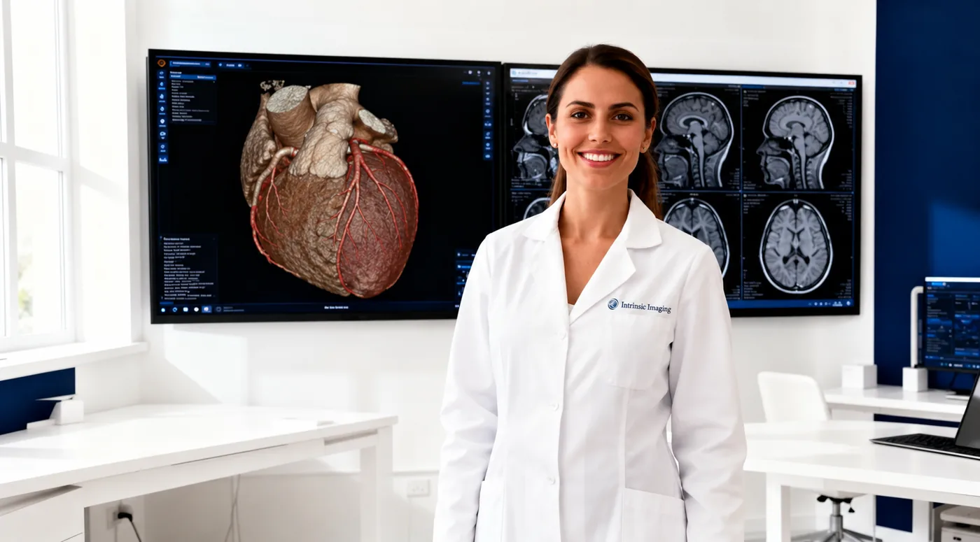
Radiologist performing blinded independent central review at Intrinsic Imaging

Definition

What an Imaging Core Lab Does

In a multicenter trial, images are acquired by different technologists on different scanners and would otherwise be interpreted by different local radiologists. An imaging core lab removes that variability. It standardizes how images are acquired, receives and quality-checks every exam centrally, and has independent, blinded readers apply the same criteria to every subject at every timepoint.

The result is an imaging endpoint that regulators can trust: measured the same way across the study, free from site-level bias, and supported by a traceable record from the scanner to the dataset. FDA guidance on clinical trial imaging endpoints describes this centralized, blinded model as the expected approach when imaging supports a primary or key secondary endpoint.

Standardized acquisition

Acquisition guidelines, scanner qualification and phantom QC mean a lesion measured in Boston and one measured in Seoul are measured on comparable images.

Independent, blinded interpretation

Subspecialty readers assess images without knowledge of treatment arm, site assessment or clinical course, following the response criteria written into the imaging charter.

A traceable record

Every intake, query, read, adjudication and data transfer is recorded in a validated, 21 CFR Part 11 compliant system, so the evidence can be reconstructed during audit or inspection.

Inside the core lab

How Our Imaging Core Lab Works

  1. Imaging charter and read design

    The charter defines the imaging schedule, acquisition parameters, response criteria, reader model, blinding rules and adjudication triggers before the first subject is scanned. It is the document every later step is checked against. Typical turnaround: charter finalized in about 20 business days after kickoff.

  2. Site qualification and acquisition standardization

    Equipment surveys, scanner validation, protocol-specific acquisition parameters, test submissions and phantom QC where the study requires it. Technologists are trained on the acquisition guidelines before routine study imaging begins. Typical turnaround: reading system ready in about 15 business days.

  3. Secure image intake and quality control

    Sites upload images through a secure, DICOM-based process. Each exam is de-identified, assigned to the correct subject, visit and modality, and checked for completeness and technical quality against the charter. Queries go back to the site with a clear description of what to correct. Typical turnaround: receipt confirmed the same business day; QC result or query within 24–48 hours of upload.

  4. Blinded independent central review

    Qualified exams are routed to a blinded, study-specific reading environment with single-, double- or triple-read configurations. Readers see only what the charter allows.

    Electronic assessment forms are built around the study's criteria, so every read captures the same measurements in the same way. Typical turnaround: eligibility and safety reads in 24–72 hours; efficacy read batches in 5–10 business days.

  5. Quantitative analysis and adjudication

    Longitudinal measurements, volumetry and imaging biomarker analyses are performed under written method specifications, with reproducibility checks. When readers disagree beyond the charter's discordance rules, an adjudicator reviews the relevant images and evidence, and the outcome is recorded with the version history intact.

  6. Data delivery and regulatory support

    Completed assessments are reconciled against source reads, mapped to agreed transfer specifications and released as validated, 21 CFR Part 11 compliant datasets, aligned to interim analyses, database lock and submission milestones. The same records support FDA, EMA and PMDA review and any inspection that follows.

Full turnaround expectations for each step

Who reads your images

Subspecialty Readers, Held to One Standard

Intrinsic Imaging draws on more than 650 board-certified, fellowship-trained physicians across Oncology, Cardiovascular, Central Nervous System, Gastroenterology, Musculoskeletal and Ophthalmology. Readers are matched to the indication, the modality and the response criteria your protocol specifies.

Before a study opens, readers are trained on the imaging charter and complete study-specific qualification cases. Reader performance, including intra- and inter-reader agreement, is monitored throughout the study, and retraining is triggered when the charter's thresholds are not met. Reader independence from the sponsor and from participating sites is documented.

How readers are qualified and monitored Meet the leadership team

Quality Management System

Every step above runs inside a quality management system certified by BSI to five ISO standards and inspected three times by the FDA with zero Form FDA 483 observations. Details are on our Quality Management System, ISO certifications and FDA inspections pages.

Software validation

Endpoints we read

Modalities and Response Criteria

Our core lab reads CT, MRI, PET/CT, SPECT, ultrasound and echocardiography, radiography, DXA, angiography, intravascular imaging, retinal imaging and central endoscopy. Established criteria include RECIST 1.1, iRECIST, mRECIST, RANO, Lugano, PCWG3, PERCIST, Choi, LVEF and strain, amyloid and tau PET, volumetric MRI, Sharp/van der Heijde, RAMRIS, MOAKS, Kellgren-Lawrence, SES-CD, Mayo endoscopic subscore, MRI-PDFF and elastography, alongside protocol-specific endpoints.

Browse therapeutic areas

Transferring from another core lab?

We take over image archives, harmonize the imaging charter and mobilize subspecialty readers so an active study keeps its timelines.

Study rescue and core lab transfer

Frequently Asked Questions

What is an imaging core lab?
An imaging core lab is a central facility that standardizes image acquisition across trial sites, receives and quality-checks every examination, and provides blinded, independent interpretation by qualified readers according to the imaging charter. Intrinsic Imaging fulfills these responsibilities to generate consistent, unbiased imaging endpoints for regulatory evaluation.
When does a trial require an imaging core lab?
Engaging an imaging core lab such as Intrinsic Imaging is warranted when imaging supports a primary or key secondary endpoint, a trial spans multiple sites or scanner types, or the design requires blinded independent central review. Engaging Intrinsic Imaging is also warranted when a medical device or AI/CAD product is evaluated against an expert reference standard.
How does Intrinsic Imaging select physicians for a study?
Intrinsic Imaging selects readers from a network of 650+ board-certified and fellowship-trained physicians according to therapeutic expertise and the study’s assessment requirements. Intrinsic Imaging provides protocol-specific training on the imaging charter, applicable criteria and documented review procedures before assessments begin.
Go to more Frequently Asked Questions

Guidance for Sponsors

Physician-authored guidance from Intrinsic Imaging on imaging endpoints and trial design.

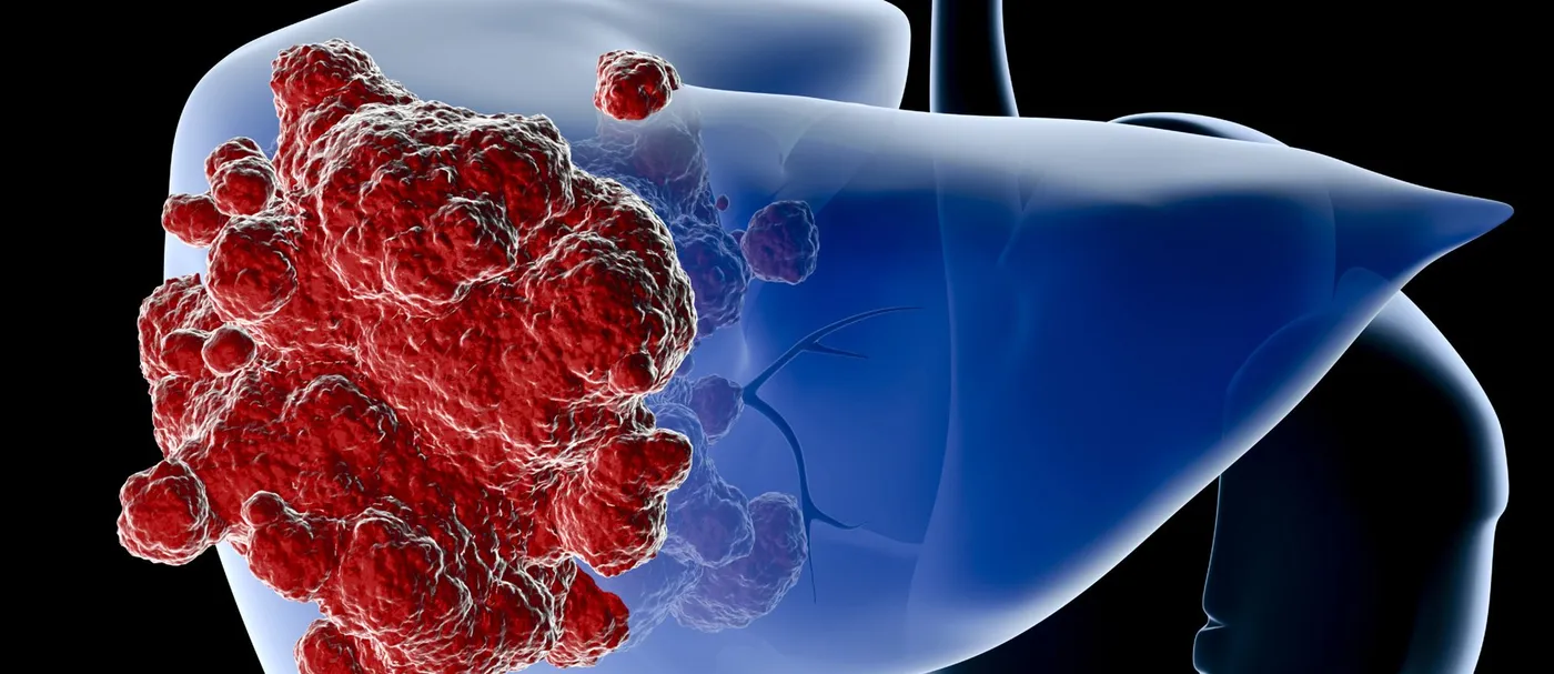
Illustration of a liver with a cluster of red tumor cells on its surface, representing imaging of tumor response

Guidance for Sponsors · Volume 6, Issue 3 · 2026 Edition

Imaging and Monitoring Tumor Response in Clinical Trials

Imaging is the way most cancer trials measure whether a drug works. Objective response rate, progression-free survival and duration of response all depend on images acquired at protocol-defined intervals and interpreted by consistent rules. Over the past fifteen years those rules have multiplied: RECIST 1.1 remains the default, but immunotherapy, molecularly targeted agents, locoregional therapies and PET have each produced their own criteria. This issue explains the current landscape of response assessment and what sponsors must build into a protocol to make imaging endpoints hold up to regulatory review.

Read the article
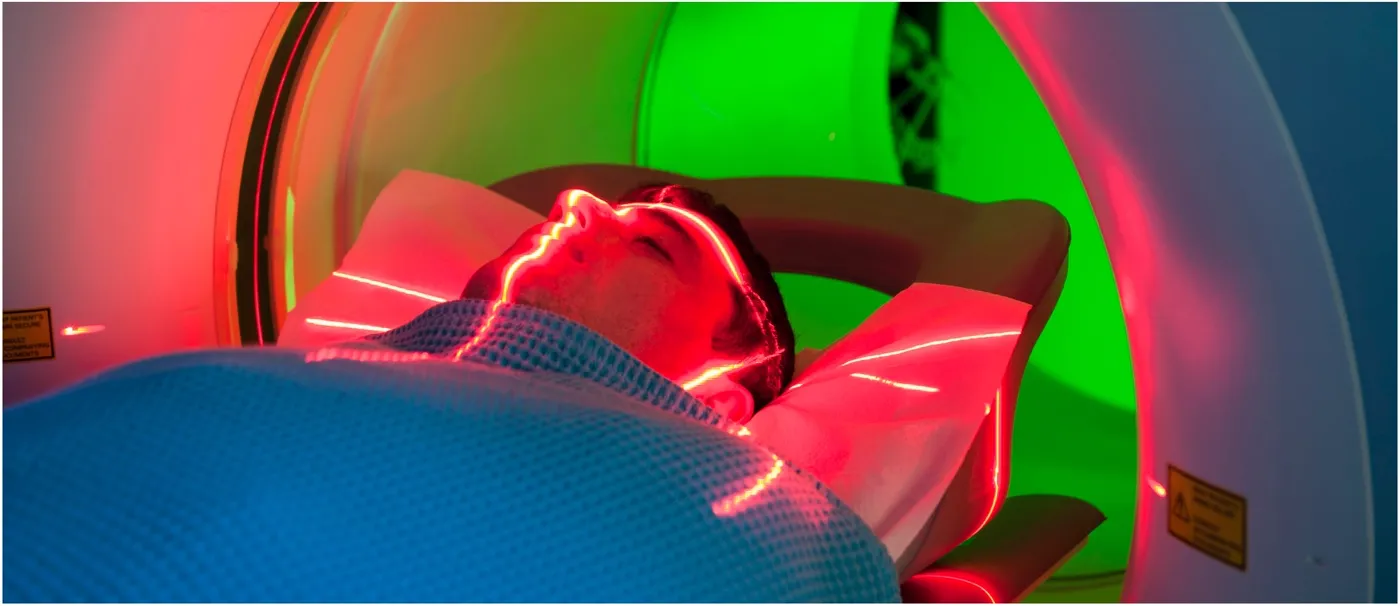
Patient positioned in a CT scanner

Guidance for Sponsors · Volume 4, Issue 7 · 2026 Edition

Computed Tomography Data Acquisition in Clinical Trials

CT is one of the most widely used modalities in clinical trials, and a substantial literature, led by RECIST 1.1, governs how lesions are measured. Far less attention is paid to how the images are acquired, yet patient motion, scanner variability, slice thickness and contrast timing all add error to every measurement. FDA's 2018 guidance on imaging endpoint process standards makes acquisition a regulatory concern. A few basic principles, written into the protocol and imaging charter and applied consistently at every site, substantially improve data accuracy.

Read the article
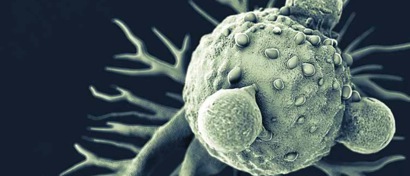
Illustration of an immune cell engaging a tumor cell

Guidance for Sponsors · Volume 5, Issue 5 · 2026 Edition

irRECIST and Immuno-Oncology in Clinical Trials

Immunotherapy has changed the practice of oncology, and it has changed the practice of measuring response. Tumors treated with checkpoint inhibitors can enlarge, or show new lesions, before they shrink, a pattern that conventional criteria classify as failure. Over fifteen years, response assessment has moved from the immune-related response criteria of 2009, through unidimensional irRC and irRECIST, to iRECIST, the RECIST Working Group's consensus standard. This issue traces that evolution, compares the criteria and explains how sponsors should apply them so that a working therapy is not mistaken for a failing one.

Read the article
Browse all Guidance for Sponsors →

Tell us about your trial.

We'll propose a read design, reader panel and turnaround plan for your imaging endpoints, and put all three in writing in the imaging charter.