
Guidance for Sponsors · Volume 5, Issue 3 · 2026 Edition
Functional Imaging of the Dopamine Pathway
By Barry Menick, M.D., Board-Certified Neuroradiologist · Updated for 2026 by Intrinsic Imaging
Download the PDF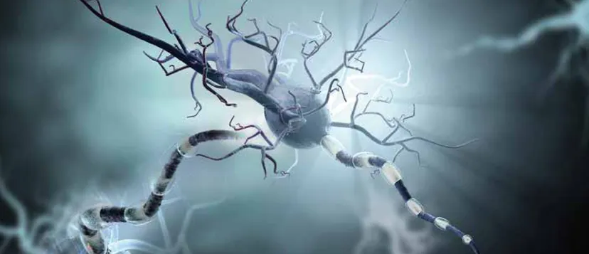
Parkinson disease is rising rapidly worldwide, and its early diagnosis remains difficult: symptoms overlap with essential tremor, drug-induced parkinsonism and the atypical parkinsonian syndromes, and routine MRI is usually normal. Functional imaging of the presynaptic dopamine pathway, with DaTscan SPECT and Fluorodopa F 18 PET, makes the loss of dopaminergic neurons visible. European regulators have qualified dopamine transporter imaging as an enrichment biomarker for early Parkinson disease trials, and it now works alongside the alpha-synuclein seed amplification assay in a new biological definition of the disease. This issue explains how these tools work and how sponsors should use them.
Neurodegenerative Disorders
The diagnosis of movement disorders and neurodegenerative diseases challenges clinicians and radiologists alike. Early presentations overlap considerably, especially when tremor is absent or atypical, and routine anatomic imaging is insensitive: early findings are usually absent or nonspecific.
After Alzheimer disease, Parkinson disease (PD) is the most common neurodegenerative disorder. An estimated 1.1 million people in the United States live with PD, a number expected to reach 1.2 million by 2030,[1] and global prevalence has doubled in 25 years to more than 8.5 million people.[2]
Idiopathic PD results from degeneration of the neurons that connect the substantia nigra of the midbrain to the corpus striatum of the basal ganglia, the nigrostriatal pathway. These neurons release dopamine to receptor cells in the striatum that govern motor function. Dopamine transporter proteins on the presynaptic membrane continuously recover dopamine from the synaptic cleft once it has acted on postsynaptic receptors. Non-degenerative parkinsonian syndromes, by contrast, may be drug-induced or vascular, the result of ischemic injury to the pathway.
Imaging the Dopamine Pathway
Two FDA-approved radiopharmaceuticals image the presynaptic dopaminergic system. DaTscan (ioflupane I 123) is used with single-photon emission computed tomography (SPECT). It binds reversibly to the dopamine transporter, which therefore serves as a surrogate marker for the integrity of nigrostriatal neurons. Approved in 2011, its labeling was expanded in November 2022 to include adults with suspected dementia with Lewy bodies.[3] Fluorodopa F 18, a fluorinated analog of L-DOPA, is used with positron emission tomography (PET). FDA approved it on October 10, 2019 to visualize dopaminergic nerve terminals in the striatum in adults with suspected parkinsonian syndromes.[4]
Circulating dopamine does not cross the blood-brain barrier, but L-DOPA is carried across by the neutral amino acid transport system and converted to dopamine by aromatic L-amino acid decarboxylase, then stored in presynaptic vesicles. DaTscan images therefore reflect transporter density in the striatum, while Fluorodopa images reflect decarboxylase activity and vesicular storage capacity in the same structures. Both studies produce images of the striatum that are interpreted by the relative intensity of uptake.
The two methods differ in practice. Fluorodopa PET offers moderately higher spatial resolution and requires less than an hour between injection and imaging, but the short half-life of fluorine-18 and the demands of synthesis have limited its use to centers with nearby cyclotrons. The iodine-123 label of DaTscan has a longer half-life, the agent is widely available, and imaging follows injection by three to four hours.
Parkinson Disease and Its Mimics
Motor symptoms of PD generally appear after 60 to 80% of dopamine-producing neurons are lost. Loss in the striatal terminals proceeds from lateral to medial, affecting the posterior putamen before the caudate head, and imaging abnormalities may be unilateral, bilateral or asymmetric. Early in PD, dopamine transporters are downregulated to preserve synaptic dopamine, which enhances the sensitivity of DaTscan; decarboxylase activity may be upregulated in compensation, so Fluorodopa PET can be falsely reassuring in early disease. Both tests reliably distinguish PD and the atypical parkinsonian syndromes, multiple system atrophy, progressive supranuclear palsy, corticobasal degeneration and dementia with Lewy bodies, from essential tremor and drug-induced parkinsonism, in which imaging is normal.
Clinical history, examination and a response to dopaminergic therapy remain the foundation of diagnosis. Under the 2015 Movement Disorder Society criteria, normal functional imaging of the presynaptic dopaminergic system is an absolute exclusion criterion for PD.[5] Yet misdiagnosis is common, particularly when PD presents without tremor or essential tremor presents asymmetrically. DaTscan SPECT is therefore a valuable safeguard against overdiagnosis and overtreatment.
Imaging in Clinical Trials
Trial experience has shown why this matters. In early PD studies, 5.7 to 14.7% of participants diagnosed by experts had scans without evidence of dopaminergic deficit (SWEDD),[6] and such subjects progress far more slowly than true PD, diluting any treatment effect. In 2018, the European Medicines Agency qualified dopamine transporter imaging as an enrichment biomarker to select patients with early motor symptoms who are likely to progress during trials of up to two years.[7]
The field is now moving toward a biological definition of disease. The cerebrospinal fluid alpha-synuclein seed amplification assay identified PD with a sensitivity of 87.7% and a specificity of 96.3% in the Parkinson's Progression Markers Initiative,[8] and in August 2024 FDA issued a Letter of Support for its use as an enrichment biomarker in clinical trials.[9] The proposed Neuronal Alpha-Synuclein Disease Integrated Staging System combines synuclein status with dopaminergic deficit on imaging to stage disease biologically.[10] Quantitative transporter imaging is also used as a progression measure: in the Phase 2 PASADENA trial of prasinezumab, putaminal dopamine transporter binding showed no substantial difference between treatment and placebo,[11] and a Phase 3 trial of the antibody in early-stage disease began in November 2025.[12]
Guidance for Sponsors
Imaging a Parkinson disease trial
- Enrich with imaging. Use dopamine transporter imaging at screening to exclude SWEDD subjects.[7]
- Pair biomarkers. Consider the seed amplification assay alongside imaging for biological enrichment.[9,10]
- Standardize acquisition. Fix the agent, dose, uptake time, camera and reconstruction across sites.
- Quantify centrally. Calculate striatal binding ratios with validated software and calibrated readers.[13]
- Prespecify endpoints. Define how imaging will support selection, staging or progression in the protocol.[13]
Summary
As the prevalence of Parkinson disease rises in an aging population, accurate documentation of disease has never been more important. Functional imaging of the dopamine pathway qualifies the right patients for clinical trials, documents disease before device implantation or targeted ablation, and now anchors a biological framework for staging. Because PD and the atypical parkinsonian syndromes can also cause cognitive decline, DaTscan SPECT and Fluorodopa PET will increasingly be used together with volumetric MRI, FDG-PET and amyloid PET to reach accurate diagnoses.
About the Author
Dr. Barry Menick, a board-certified neuroradiologist, wrote the original edition of this issue at Intrinsic Imaging. He earned his undergraduate and medical degrees at Duke University and completed his residency in diagnostic radiology and a two-year neuroradiology fellowship at the Hospital of the University of Pennsylvania. A Senior Member of the American Society of Neuroradiology since 1991 and a member of the American Society of Functional Neuroradiology, he has a particular interest in quantitative imaging of neurodegenerative disease and functional imaging of brain tumors.
References
- Parkinson's Foundation. Parkinson's disease statistics. https://www.parkinson.org/understanding-parkinsons/statistics
- World Health Organization. Parkinson disease fact sheet. https://www.who.int/news-room/fact-sheets/detail/parkinson-disease
- GE HealthCare. DaTscan (ioflupane I 123 injection) indication expanded to include use in patients with suspected dementia with Lewy bodies. November 3, 2022. https://www.gehealthcare.com/en/about/newsroom/press-releases/datscan-indication-expanded-to-include-use-in-patients-with-suspected-dementia-with-lewy-bodies
- U.S. FDA. Drug trials snapshot: Fluorodopa F 18. Approved October 10, 2019. https://www.fda.gov/drugs/drug-approvals-and-databases/drug-trials-snapshots-fluorodopa-f-18
- Postuma RB, Berg D, Stern M, et al. MDS clinical diagnostic criteria for Parkinson's disease. Mov Disord. 2015;30(12):1591–1601. https://www.med.upenn.edu/digitalneuropathologylab/assets/user-content/documents/educational-resources/mds-clinical-diagnostic-criteria-for-parkinson%27s-disease.pdf
- Stockner H, Schwingenschuh P, Djamshidian A, et al. Is transcranial sonography useful to distinguish scans without evidence of dopaminergic deficit patients from Parkinson's disease? Mov Disord. 2012;27(9):1182–1185. https://pmc.ncbi.nlm.nih.gov/articles/PMC3660780/
- European Medicines Agency, CHMP. Qualification opinion on dopamine transporter imaging as an enrichment biomarker for Parkinson's disease clinical trials in patients with early Parkinsonian symptoms. May 29, 2018. https://www.ema.europa.eu/en/documents/regulatory-procedural-guideline/qualification-opinion-dopamine-transporter-imaging-enrichment-biomarker-parkinsons-disease-clinical-trials-patients-early-parkinsonian-symptoms_en.pdf
- Siderowf A, Concha-Marambio L, Lafontant DE, et al. Assessment of heterogeneity among participants in the Parkinson's Progression Markers Initiative cohort using α-synuclein seed amplification: a cross-sectional study. Lancet Neurol. 2023;22(5):407–417. https://pubmed.ncbi.nlm.nih.gov/37059509/
- U.S. FDA. Drug development tool letter of support DDT-BMQ-000157 to the Critical Path for Parkinson's consortium. August 19, 2024. https://www.fda.gov/media/181368/download
- Simuni T, Chahine LM, Poston K, et al. A biological definition of neuronal α-synuclein disease: towards an integrated staging system for research. Lancet Neurol. 2024;23(2):178–190. https://pubmed.ncbi.nlm.nih.gov/38267190/
- Pagano G, Taylor KI, Anzures-Cabrera J, et al. Trial of prasinezumab in early-stage Parkinson's disease. N Engl J Med. 2022;387(5):421–432. https://pubmed.ncbi.nlm.nih.gov/35921451/
- ClinicalTrials.gov. NCT07174310: A study to evaluate the efficacy and safety of intravenous prasinezumab in participants with early-stage Parkinson's disease. https://clinicaltrials.gov/study/NCT07174310
- U.S. FDA. Clinical trial imaging endpoint process standards: guidance for industry. April 2018. https://www.fda.gov/regulatory-information/search-fda-guidance-documents/clinical-trial-imaging-endpoint-process-standards-guidance-industry
Planning a Parkinson disease trial?
Talk with our team about dopamine transporter imaging, quantitative striatal analysis and independent neuroradiology review.
Contact UsRelated reading: Temporal Lobe Epilepsy and Volumetric MRI in Clinical Trials · Muscular Dystrophy: Medical Imaging in Clinical Trials · Oncologic Imaging: The Benefits of Diffusion-Weighted MRI