
Guidance for Sponsors · Volume 4, Issue 2 · 2026 Edition
Screening Mammography AI: A Continued Market Opportunity
By Richard Benedikt, M.D., MBA, Board Certified Radiologist · Updated for 2026 by Intrinsic Imaging
Download the PDF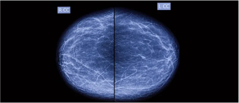
Computer-aided detection (CAD) promised to help radiologists find more breast cancers, but a decade of large studies showed that first-generation systems added false alarms without improving accuracy. Artificial intelligence has changed that picture. Randomized and nationwide studies published since 2023 show AI-supported screening detecting more cancers, reducing interval cancers and cutting radiologist workload without raising recall rates. For developers, the opportunity is larger than ever. So is the burden of proof: regulators, payers and screening programs now expect rigorous reader studies and real-world evidence.
Screening Mammography and CAD
Screening mammography remains the foundation of breast cancer early detection. In April 2024, the U.S. Preventive Services Task Force updated its recommendation to biennial screening mammography for all women aged 40 to 74.[1] The technology has changed considerably: as of September 2026, 8,657 of the 9,128 FDA-certified mammography facilities in the United States operate digital breast tomosynthesis (DBT) units, with 13,575 accredited DBT units in service.[2]
Computer-aided detection has a long history in this setting. The FDA approved the first mammography CAD system in 1998, and the technology spread rapidly into practice.[3] Early commercial products were designed for analog systems: film mammograms were digitized with a laser scanner and analyzed by proprietary software, which placed one type of mark on calcifications and another on masses or areas of increased density. With the conversion to full-field digital mammography and then to DBT, CAD marks moved to soft-copy display alongside the radiologist's review.
The rationale has not changed. Screening mammography misses a meaningful share of cancers, and errors in interpretation fall broadly into three categories: perception, analysis and technical. CAD was designed to address perception, prompting the radiologist to look again at findings that might otherwise be overlooked, while leaving the analysis of their significance to the radiologist.
Lessons from First-Generation CAD
The clinical results of traditional CAD were controversial from the start. Early investigators reported improvements in the number of screen-detected cancers and a greater proportion of early-stage cancers. Larger studies told a different story. In 2007, Fenton and colleagues reported in the New England Journal of Medicine that CAD use at community facilities was associated with reduced specificity and more biopsies, without a clear improvement in cancer detection.[3] In 2015, Lehman and colleagues analyzed digital screening mammograms in the Breast Cancer Surveillance Consortium and found that screening performance was not improved with CAD on any metric assessed. Sensitivity was 85.3% with CAD and 87.3% without it, even though CAD was used for most U.S. screening mammograms at a cost of more than $400 million a year.[4]
A review of these studies shows why results varied. Technical factors (analog versus digital, the number of views, the algorithm itself), professional factors (radiologist and technologist experience) and study design (retrospective versus prospective, cancer-enriched versus unselected populations) all influenced the outcome. Traditional CAD also generated many marks per examination, most of them benign, placing a heavy burden on the radiologist. Many practitioners reported a rise in call-back rates after implementing CAD. Call-backs matter: they add cost, radiation exposure and patient anxiety, they are monitored against national guidelines, and they are central to any regulatory submission.
The AI Generation
Two developments reopened the market. First, regulation changed. In January 2020, the FDA reclassified computer-assisted detection devices for mammography from Class III to Class II, moving them from the premarket approval pathway to 510(k) clearance with special controls.[5] Second, deep-learning algorithms began to deliver the gains that traditional CAD did not.
The evidence is now prospective and large. In the Swedish MASAI randomized trial of more than 105,000 women, AI-supported screening detected 6.4 cancers per 1,000 women, compared with 5.0 with standard double reading, while reducing screen-reading workload by 44.3%.[6] Final results published in The Lancet in January 2026 reported a 12% lower interval cancer rate, sensitivity of 80.5% versus 73.8%, and identical specificity of 98.5% in both groups.[7] In Germany, the PRAIM implementation study of 461,818 women read by 119 radiologists found a 17.6% higher cancer detection rate with AI (6.70 versus 5.70 per 1,000) and no increase in recall (37.4 versus 38.3 per 1,000).[8]
The lesson for developers is clear. AI succeeds when it is validated in the way radiologists actually read, at the scale screening programs actually operate, and with the endpoints that matter: cancer detection, interval cancers, recall and workload.
Guidance for Sponsors
Designing a mammography AI study
- Plan the reader study carefully. Multi-reader, multi-case (MRMC) studies are central to clearance. Define reader number and experience, case enrichment, washout periods and reading conditions in advance, following FDA's guidance on clinical performance assessment of CAD devices.[5]
- Cover the modalities in use. With DBT now in nearly every certified U.S. facility, include DBT as well as 2D digital mammography, and images from multiple vendors.[2]
- Establish rigorous ground truth. Confirm cancers with pathology and negatives with adequate follow-up, so that interval cancers are captured.
- Measure recall and specificity, not only sensitivity. The history of CAD shows that detection gains alone do not persuade regulators or clinicians.
- Report results by breast density. Since September 10, 2024, U.S. facilities must notify patients of their breast density, and performance in dense breasts is a question every buyer will ask.[9]
- Reflect real workflow. Test how radiologists interact with AI outputs, not only standalone algorithm performance.
Summary
Screening mammography remains the most widely used tool for reducing breast cancer deaths, and the case for computer assistance has never been stronger. First-generation CAD taught the field that more marks do not equal better medicine. The current generation of AI has shown, in randomized and nationwide studies, that it can find more cancers with fewer false alarms and less radiologist time. A significant market opportunity remains for developers whose algorithms can prove that value through independent, well-designed clinical evidence.
About the Author
Dr. Richard Benedikt, a board-certified radiologist, wrote the original edition of this issue as Medical Director, Breast Imaging, at Intrinsic Imaging. He completed his diagnostic radiology residency at Walter Reed Army Medical Center, where he was chief resident, and served as Chief of Thoracic Imaging and Chief of Ultrasound at Brooke Army Medical Center. He was previously a research fellow for two years at McLean Hospital (Harvard Medical School). At the time of the original edition, he had participated in clinical trials for more than 15 years, had authored or coauthored more than 25 publications, and served as a manuscript reviewer for two major radiology journals, AJR and JACR. Dr. Benedikt also holds an MBA from the University of Texas.
References
- U.S. Preventive Services Task Force. Breast cancer: screening. April 30, 2024. https://www.uspreventiveservicestaskforce.org/uspstf/recommendation/breast-cancer-screening
- U.S. FDA. MQSA 2026 scorecard statistics. September 2026. https://www.fda.gov/radiation-emitting-products/mqsa-insights/2026-scorecard-statistics
- Fenton JJ, Taplin SH, Carney PA, et al. Influence of computer-aided detection on performance of screening mammography. N Engl J Med. 2007;356(14):1399–1409. https://pmc.ncbi.nlm.nih.gov/articles/PMC3182841/
- Lehman CD, Wellman RD, Buist DSM, et al. Diagnostic accuracy of digital screening mammography with and without computer-aided detection. JAMA Intern Med. 2015;175(11):1828–1837. https://pubmed.ncbi.nlm.nih.gov/26414882/
- U.S. FDA. Clinical performance assessment: considerations for computer-assisted detection devices applied to radiology images and radiology device data in premarket notification (510(k)) submissions. https://www.fda.gov/media/77642/download
- Hernström V, et al. Screening performance and characteristics of breast cancer detected in the MASAI trial. Lancet Digit Health. 2025. https://www.thelancet.com/journals/landig/article/PIIS2589-7500(25)00004-4/fulltext
- The Lancet publishes final results from the first randomized controlled trial in breast AI (MASAI). January 30, 2026. https://www.prnewswire.com/news-releases/the-lancet-publishes-final-results-from-the-first-randomized-controlled-trial-in-breast-ai-302675094.html
- Eisemann N, et al. Nationwide real-world implementation of AI for cancer detection in population-based mammography screening. Nat Med. 2025;31(3):917–924. https://www.nature.com/articles/s41591-024-03408-6
- U.S. FDA. Important information: final rule to amend the Mammography Quality Standards Act. https://www.fda.gov/radiation-emitting-products/mammography-quality-standards-act-mqsa-and-mqsa-program/important-information-final-rule-amend-mammography-quality-standards-act-mqsa
Developing a mammography AI or CAD device?
Talk with our team about MRMC reader study design, DBT and 2D case sets, and FDA-ready clinical evidence.
Contact UsRelated reading: Magnetic Resonance Elastography: A Quantitative Endpoint for Liver Fibrosis Trials · Oncologic Imaging: The Benefits of Diffusion-Weighted MRI · Imaging Analysis and Operative Treatment of Diaphyseal Fractures