
Guidance for Sponsors · Volume 4, Issue 4 · 2026 Edition
Imaging Analysis and Operative Treatment of Diaphyseal Fractures
By Jack Clement, M.D., Ph.D., Medical Director, Musculoskeletal Imaging · Updated for 2026 by Intrinsic Imaging
Download the PDF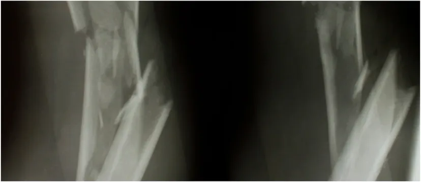
Fractures are among the most common reasons for an imaging examination, and imaging guides every stage of care: diagnosis, classification, reduction, fixation, detection of complications and confirmation of healing. Radiography, CT, MRI and bone scintigraphy each play a distinct role. For sponsors of orthopedic drug, biologic and device trials, the central challenge is that fracture union, the most commonly reported primary outcome, still has no universally accepted radiographic definition. Standardized scoring, consistent imaging schedules and independent central review turn a subjective judgment into a defensible endpoint.
Imaging Analysis of Diaphyseal Fractures
Fracture imaging covers a broad range of questions: initial diagnosis, fragment displacement and alignment, predisposing lytic lesions, alignment after closed reduction, alignment and hardware complications after open reduction, possible infection, and the progress of healing. Each modality contributes something different.
Radiography remains the mainstay at every stage. It provides an overall assessment of fracture morphology and shows how a fracture evolves over time. At least two views are required, because a fracture invisible on one view may be obvious on another, and different projections reveal displaced fragments, subtle bone destruction, malunion or hardware failure. Because radiographs superimpose anatomy, however, they are limited when detailed, precise assessment is needed.
Computed tomography acquires images by rotating a narrow x-ray beam and an opposing detector rapidly around the patient, measuring differences in attenuation and reconstructing the image mathematically. Musculoskeletal protocols typically use 1 to 3 mm collimation for small body parts such as the hands and feet, and 3 to 5 mm for larger regions. The helical data set can be reconstructed in any plane, and bone or soft-tissue filtering algorithms are applied according to the clinical question. Compared with radiography, CT is more detailed and therefore more sensitive and specific.
Bone scintigraphy is highly sensitive for occult fractures but lacks specificity. A normal bone scan virtually excludes fracture, whereas a positive scan may reflect fracture, infection, tumor or arthritis, and must be interpreted with clinical and radiographic data. Because uptake declines as bone turnover normalizes, scintigraphy can also track healing.
MRI is exquisitely sensitive to bone marrow edema, an invariable finding in fracture, and detects nondisplaced or incomplete fractures that radiography and CT miss. It may not readily distinguish bone contusion from a true cortical fracture, particularly in avulsion and small chip fractures, which CT delineates better. MRI also shows concomitant soft-tissue injury, often more important than the fracture itself. After fixation, metal artifact reduction sequences, now including multispectral techniques such as MAVRIC and SEMAC, limit distortion from hardware and allow assessment of the bone–implant interface and adjacent soft tissue.[1]
Fracture Mechanism and Classification
Diaphyseal fractures of the femur, tibia, fibula, humerus, radius, ulna and the long bones of the hands and feet are common consequences of trauma. Indirect rotational forces produce spiral or spiral wedge fractures with a large surface of bone contact and relatively little soft-tissue and periosteal damage, which favor healing. Bending forces produce oblique wedge fractures with more soft-tissue and periosteal injury. Direct forces produce transverse, fragmented wedge and complex fractures, often open, with extensive soft-tissue damage; these are the most difficult to heal.
Fracture type predicts the extent of soft-tissue injury and guides the timing of surgery and the choice of reduction and fixation technique. The goal is restoration of bone length and of axial and rotational alignment relative to the adjacent joints. In clinical trials, the AO/OTA Fracture and Dislocation Classification Compendium, last revised in 2018, provides the common language for fracture morphology and is the standard basis for eligibility criteria and stratification.[2]
Operative Treatment
Diaphyseal fractures are difficult to stabilize by nonoperative means such as casting or traction, which risk angular deformity, nonunion, shortening and stiffness of adjacent joints. Internal or external fixation usually produces better function. The aim is not anatomic reduction and rigid fixation, but adequate reduction and stable fixation: anatomic reduction often damages periosteum and soft tissue, and rigid fixation prevents the small amount of motion that stimulates healing. Every technique should stabilize the fracture while remaining as minimally invasive as possible, preserving blood supply and periosteal integrity.
Intramedullary nails act as internal splints within the medullary canal. They permit early weight bearing and micromotion at the fracture site, promoting callus formation, with proximal or distal interlocking screws to control rotation. Plates and screws are applied to the bone surface and are commonly used when a diaphyseal fracture extends into the metaphysis, often with an interfragmentary lag screw. Conventional plates compress the cortex and can compromise periosteal blood supply. Locking compression plates avoid this: the locking-head screws engage plate and bone separately, leaving a space between them while stabilizing the fracture. External fixation is used when soft-tissue damage is extensive. Because healing may be delayed and the risk of infection and loosening is higher, it generally serves as a bridge to internal fixation once the soft tissues have recovered.
Measuring Healing in Clinical Trials
Bone union is the most commonly reported primary outcome in fracture trials, yet no universally accepted radiographic definition exists. A 2026 review found that the familiar rule of bridging callus on three of four cortices has no traceable validation study, and that among 123 clinical studies, 11 different radiographic criteria were used and only two reported a quantitative measure of interobserver reliability.[3]
Standardized scores address this problem. The Radiographic Union Scale in Tibial fractures (RUST) scores callus and fracture-line visibility at each of four cortices, giving a total of 4 (not healed) to 12 (healed). Independent validation reported an intraclass correlation of 0.75 for individual readers, rising to 0.94 when scores from several readers were averaged.[4] The modified RUST (mRUST) adds finer gradation and is now widely used, including for femoral shaft fractures.[5] Where radiographs are equivocal, CT improves agreement: in one study of clavicle fractures, interobserver agreement on union was 0.75 with CT versus 0.50 with radiographs.[6]
Guidance for Sponsors
Designing a fracture-healing endpoint
- Define union in the protocol. State the scoring system (RUST or mRUST), the threshold for union and the views required, rather than relying on an undefined clinical judgment.[3,4]
- Classify consistently. Use the AO/OTA compendium for eligibility and stratification, confirmed by the central reader.[2]
- Standardize the imaging schedule. Fix time points, projections and positioning, and include the immediate postoperative baseline, which improves scoring reliability.[4]
- Use CT selectively. Reserve CT for equivocal or delayed union, or as a confirmatory endpoint, with a dose-conscious protocol.[6]
- Plan for hardware. Specify metal artifact reduction MRI when soft tissue, infection or the bone–implant interface is an endpoint.[1]
- Read centrally with more than one reader. Blinded independent review with adjudication reduces variability; averaged scores from multiple readers are markedly more reliable.[4]
Summary
Imaging is central to fracture care at every stage of healing. Radiography provides the overall assessment of morphology and complications; CT adds exquisite bone detail for fragment alignment and callus formation; MRI detects occult fractures and the soft-tissue injury that often guides therapy. Current research combines novel devices, biologics and surgical approaches to achieve adequate reduction and stable fixation with minimal tissue damage. For sponsors, demonstrating that benefit depends on a precisely defined, reliably measured and independently read imaging endpoint.
About the Author
Dr. Jack Clement wrote the original edition of this issue as Medical Director, Musculoskeletal Imaging, at Intrinsic Imaging. A graduate of Tulane University in New Orleans, he completed M.D. and Ph.D. degrees at Baylor College of Medicine in Houston, where his training was sponsored by the National Institutes of Health. He has coauthored peer-reviewed original research in Science, the New England Journal of Medicine and Neuron. He completed a diagnostic radiology residency at Baylor College of Medicine and subspecialized in musculoskeletal radiology at Harvard Medical School's Massachusetts General Hospital in Boston. He is a board-certified radiologist with specialized training in orthopedic radiology and interventional pain management.
References
- Managing hardware-related metal artifacts in MRI: current and future approaches. Skeletal Radiol. 2024. https://pmc.ncbi.nlm.nih.gov/articles/PMC11303499/
- Meinberg EG, Agel J, Roberts CS, Karam MD, Kellam JF. Fracture and Dislocation Classification Compendium—2018. J Orthop Trauma. 2018;32 Suppl 1:S1–S170. https://pubmed.ncbi.nlm.nih.gov/29256945/
- Koh JH, Sakong S. How do we know a fracture has healed? A narrative review of radiographic bone union definitions and assessment methods. J Musculoskelet Trauma. 2026;39(3):185–194. https://e-jmt.org/journal/view.php?number=2411
- Leow JM, Clement ND, Tawonsawatruk T, Simpson CJ, Simpson AHRW. The radiographic union scale in tibial (RUST) fractures: reliability of the outcome measure at an independent centre. Bone Joint Res. 2016;5(4):116–121. https://boneandjoint.org.uk/Article/10.1302/2046-3758.54.2000628
- Reliability of modified Radiographic Union Score for Tibia (mRUST) in diaphyseal femur fractures. JAAOS Glob Res Rev. 2022. https://journals.lww.com/jaaosglobal/fulltext/2022/05000/reliability_of_modified_radiographic_union_score.11.aspx
- The accuracy of computed tomography for clavicle non-union evaluation. https://pmc.ncbi.nlm.nih.gov/articles/PMC8039767/
Planning an orthopedic or fracture-healing trial?
Talk with our team about union endpoints, RUST and mRUST scoring, imaging schedules and independent central review.
Contact UsRelated reading: Magnetic Resonance Elastography: A Quantitative Endpoint for Liver Fibrosis Trials · Screening Mammography AI: A Continued Market Opportunity · Oncologic Imaging: The Benefits of Diffusion-Weighted MRI