Insights and Publications

Guidance for Sponsors and other publications from Intrinsic Imaging physicians on imaging endpoints, core lab methods and clinical trial design.

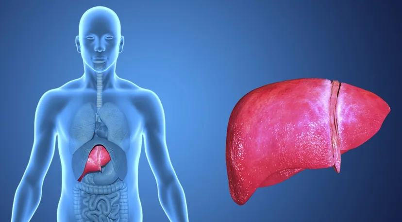
Illustration of the human liver

Guidance for Sponsors · Volume 4, Issue 1 · 2026 Edition

Magnetic Resonance Elastography: A Quantitative Endpoint for Liver Fibrosis Trials

Magnetic resonance elastography (MRE) measures liver stiffness without a needle, and it has become one of the most accurate imaging tools available for staging liver fibrosis. Now that the FDA has approved the first therapies for metabolic dysfunction-associated steatohepatitis (MASH), sponsors developing the next generation of liver therapies need endpoints that are quantitative, repeatable and acceptable to patients. Combined with MRI proton density fat fraction (MRI-PDFF) in a single liver MRI exam, MRE provides exactly that: a one-minute acquisition that stages fibrosis and tracks how it changes over the course of a trial.

Read the article
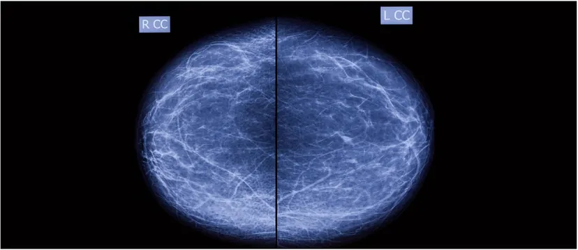
Bilateral craniocaudal screening mammogram

Guidance for Sponsors · Volume 4, Issue 2 · 2026 Edition

Screening Mammography AI: A Continued Market Opportunity

Computer-aided detection (CAD) promised to help radiologists find more breast cancers, but a decade of large studies showed that first-generation systems added false alarms without improving accuracy. Artificial intelligence has changed that picture. Randomized and nationwide studies published since 2023 show AI-supported screening detecting more cancers, reducing interval cancers and cutting radiologist workload without raising recall rates. For developers, the opportunity is larger than ever. So is the burden of proof: regulators, payers and screening programs now expect rigorous reader studies and real-world evidence.

Read the article
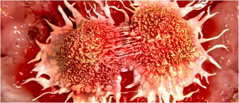
Illustration of dividing cancer cells

Guidance for Sponsors · Volume 4, Issue 3 · 2026 Edition

Oncologic Imaging: The Benefits of Diffusion-Weighted MRI

Diffusion-weighted MRI (DWI) was first used to greatest advantage in stroke imaging. Advances in gradients and pulse sequences have since made it a core tool for detecting, characterizing and following malignant lesions in the chest, abdomen and pelvis. Today it is the dominant sequence for peripheral-zone prostate cancer, a foundation of whole-body MRI for bone and marrow disease, and, through the apparent diffusion coefficient (ADC), a quantitative biomarker with published repeatability standards. For sponsors, DWI offers a fast, contrast-free measure of tumor cellularity and treatment response, provided acquisition and analysis are standardized across sites.

Read the article
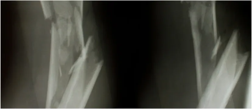
Radiographs of a comminuted long-bone diaphyseal fracture

Guidance for Sponsors · Volume 4, Issue 4 · 2026 Edition

Imaging Analysis and Operative Treatment of Diaphyseal Fractures

Fractures are among the most common reasons for an imaging examination, and imaging guides every stage of care: diagnosis, classification, reduction, fixation, detection of complications and confirmation of healing. Radiography, CT, MRI and bone scintigraphy each play a distinct role. For sponsors of orthopedic drug, biologic and device trials, the central challenge is that fracture union, the most commonly reported primary outcome, still has no universally accepted radiographic definition. Standardized scoring, consistent imaging schedules and independent central review turn a subjective judgment into a defensible endpoint.

Read the article
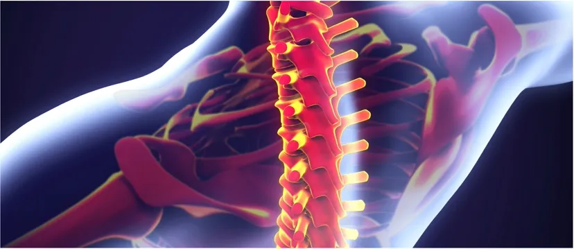
Illustration of the human spine and skeleton

Guidance for Sponsors · Volume 4, Issue 5 · 2026 Edition

Orthopedic Device Assessments: Medical Imaging Challenges in Clinical Trials

Orthopedic research spans joint reconstruction, spinal fusion and motion preservation, and biologics for cartilage, tendon and ligament repair. Regulators and payers expect rigorous quantitative and qualitative evidence of efficacy, safety and longevity, and imaging supplies much of it. Because investigational devices are often unlike anything imaged before, protocols must be built around the device itself: its engineering, materials, surgical technique and position. Advances in CT, metal-artifact-reduction MRI and radiostereometric analysis now allow earlier and more precise answers than ever.

Read the article
Illustration of the lungs and airways

Guidance for Sponsors · Volume 4, Issue 6 · 2026 Edition

The Challenges of Reducing Lung Cancer Mortality with Screening

Lung cancer remains the leading cause of cancer death in the United States. Two large randomized trials, NLST and NELSON, have shown that low-dose CT (LDCT) screening reduces lung cancer mortality, and U.S. eligibility was broadened in 2021. Yet the challenges that shaped the first edition of this issue remain: false positives, overdiagnosis, radiation, and the biases that can make screening look more effective than it is. Artificial intelligence for nodule detection and characterization adds new opportunity, and new questions that only well-designed trials can answer.

Read the article
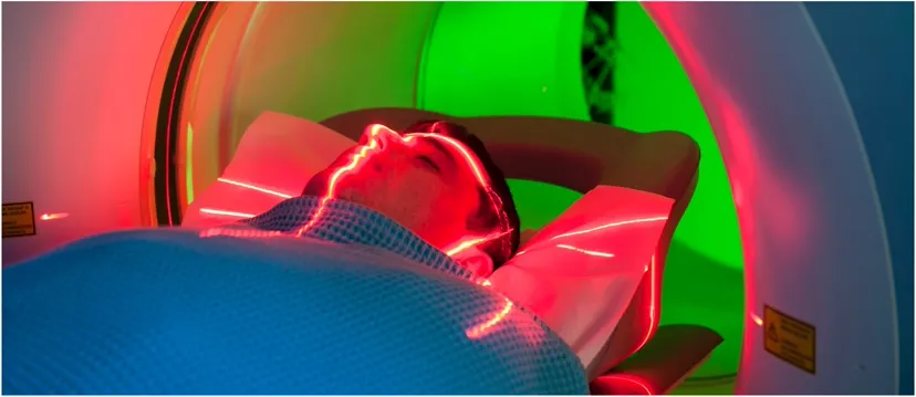
Patient positioned in a CT scanner

Guidance for Sponsors · Volume 4, Issue 7 · 2026 Edition

Computed Tomography Data Acquisition in Clinical Trials

CT is one of the most widely used modalities in clinical trials, and a substantial literature, led by RECIST 1.1, governs how lesions are measured. Far less attention is paid to how the images are acquired, yet patient motion, scanner variability, slice thickness and contrast timing all add error to every measurement. FDA's 2018 guidance on imaging endpoint process standards makes acquisition a regulatory concern. A few basic principles, written into the protocol and imaging charter and applied consistently at every site, substantially improve data accuracy.

Read the article
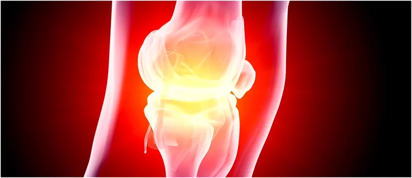
Illustration of an inflamed knee joint

Guidance for Sponsors · Volume 4, Issue 8 · 2026 Edition

Advances in High-Field MRI for the Assessment of Arthritis

Arthritis is a leading cause of disability, affecting about 53 million U.S. adults. Its central feature is loss of hyaline cartilage, which has little capacity to repair. Radiographs show only late damage; high-field 3.0 Tesla MRI shows cartilage morphology in fine detail, and compositional techniques such as T2 mapping reveal degeneration before visible loss. For sponsors, the challenge is regulatory as well as technical: FDA has not yet accepted a structural endpoint for osteoarthritis approval, so imaging must be precise, reproducible and linked to how patients feel and function.

Read the article
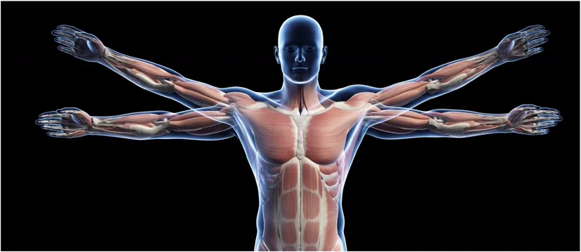
Illustration of the human muscular system

Guidance for Sponsors · Volume 4, Issue 9 · 2026 Edition

Muscular Dystrophy: Medical Imaging in Clinical Trials

MRI has become one of the most valuable tools for measuring disease in muscular dystrophy. It shows which muscles are involved, how severely and how actively, and it can guide biopsy and monitor therapy. Since the first edition of this issue, quantitative MRI has moved from grading scales to precise fat-fraction measurement, and muscle fat fraction has been shown to predict loss of ambulation in Duchenne muscular dystrophy. With new therapies now approved, MRI is a key secondary endpoint and a candidate surrogate for future trials.

Read the article
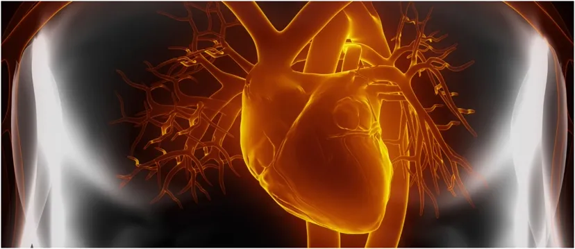
Illustration of the heart and coronary vessels

Guidance for Sponsors · Volume 4, Issue 10 · 2026 Edition

Echocardiography: Advantages in Clinical Trials

Echocardiography is safe, portable, affordable and available worldwide, which makes it one of the most practical imaging tools for clinical trials. It measures chamber size, function, valves and mass, and changes in these measures predict major cardiovascular events, allowing smaller trials and earlier answers. Since the first edition of this issue, global longitudinal strain and three-dimensional echocardiography have become standard measures, and cardio-oncology guidelines now define cardiac injury from cancer therapy partly by strain. Standardized acquisition and core-lab reading remain essential.

Read the article
Illustration of a vertebral compression fracture

Guidance for Sponsors · Volume 4, Issue 11 · 2026 Edition

Osteoporosis Therapy: Challenges in Monitoring Efficacy in Clinical Trials

Osteoporotic fractures are devastating, and proving that a new therapy prevents them has traditionally required large, long fracture trials. That changed in December 2025, when FDA qualified the 24-month change in total hip bone mineral density (BMD), measured by DXA, as a validated surrogate endpoint for phase 3 trials in postmenopausal osteoporosis. The decision makes imaging quality more important than ever. DXA must be calibrated and centrally reviewed, while QCT, vertebral fracture assessment and microarchitecture imaging add information that BMD alone cannot provide.

Read the article